
Total endovascular aortic arch repair: is it for everyone and where is its evidence?
 0
0 , ...
, ... Abstract
Open total arch replacement (TAR) remains the mainstay management strategy for thoracic aortic diseases involving the aortic arch. TAR evolved from the 2-stage conventional elephant trunk (CET) technique to the hybrid frozen elephant trunk (FET) which combined open surgical repair (OSR) with thoracic endovascular aortic repair (TEVAR) into a 1-stage procedure. Although FET has been able to achieve superior results to CET, including excellent survival, it still carries a risk of certain complications that may even require secondary reintervention. The era of elephant trunk is being overtaken by the new generation of TEVAR devices being used for total endovascular aortic arch (or endoarch) repair. Total endoarch repair (TER) is currently indicated in patients deemed high-risk for open surgery; however, it has shown strong potential for becoming the gold stand treatment for aortic arch pathologies. Despite the minimally-invasive nature of TER providing an obvious advantage over OSR in certain cases, TER remains associated with comparable mortality rates and key complications such as technical failure, neurological injury, need for reintervention, and loss of or failure to achieve target vessel patency. Upon comprehensively searching the literature, the technical success of TER ranged from 91%-100%, mortality 0%-19%, stroke 0%-16.7% and reintervention 0%-30.3%, using different commercially available endografts. Given its novelty, further studies with larger cohorts and longer follow-up periods are necessary to solidify the evidence on TER, taking into account the significant learning curve associated with TEVAR. In addition, studies directly comparing arch OSR to TER are warranted to determine superiority. This review aimed to highlight the evolution of aortic arch repair, focusing on TER device development, intervention criteria and clinical outcomes.
Keywords
INTRODUCTION
Open total arch replacement (TAR) remains the gold standard surgical approach for thoracic aortic pathologies involving the aortic arch. The reported mortality and morbidity for elective aortic arch repair are highly variable. Culpable to this overwhelming morbidity and mortality variations are correlated to patients undergoing cardiopulmonary bypass (CPB) and hypothermic circulatory arrest (HCA), in addition to risks associated with general anaesthesia. The main two techniques for TAR are the conventional elephant trunk (CET) and frozen elephant trunk (FET)[1].
Over the past decade, the thoracic endovascular approach for aortic arch (or endoarch) repair gained momentum, especially in high-risk population groups, thanks to the innovation and application instrumented by device technology endograft suppliers and the enthusiasm of endovascular surgeons. As such, endografts became available for investigational purposes in the aortic arch profile and as part of investigational device exemption programs. To this effect, certain devices are supplied fenestrated or scalloped, while others are branched, albeit single-, double-, or triple-branched stent-grafts. Such technology optimized options for aortic arch repair in high-risk patients who were deemed inoperable, which decreases the associated risk of perioperative mortality and morbidity[2,3]. However, reported series on the use of the endovascular approach in aortic arch profile continued to encounter relatively high complication rates and poor operative outcomes[4]. This can be partly attributed to revascularization requirement as well as the substantial risk of stroke due to wire and device manipulation within the aortic arch aneurysm, which is a drawback[5]. In addition, this can also be attributed to the high-risk patient population treated. Graft patency, re-intervention rates, long-term comparative functionality, and durability of endoarch were uncertain[2,4,5]. Additionally, surgeon volume-outcome linearity, learning curve, and decision-making were key factors for total endoarch repair (TER) to be considered sustainable.
In this review, we sought to highlight the TER approach, including device evolution, intervention criteria and clinical outcomes, and set the scene in a comparative mode to open surgical aortic arch repair as well as dwell on the current trend of hybridized approaches using FET which evolved from CET.
METHODS
A comprehensive literature search was performed using major search engines (PubMed, Google Scholar, EMBASE and Scopus) to search all scientific articles published as of July 2022. The search terms used included: “Aortic Dissection”, “Aortic Aneurysm”, “Conventional Elephant Trunk (OR CET)”, “Frozen Elephant Trunk (OR FET)”, “Thoracic Aorta”, “Aortic Arch”, “Endovascular”, “Endovascular Arch Repair”. Additional sources were identified by individually reviewing reference lists of included publications.
Past to present: an overview of aortic arch surgical repair
Conventional vs. frozen elephant trunk
FET and CET (evaluated in Table 1) are similar in terms of the scope of repair of the ascending aorta and the aortic arch. In both approaches, the entire transverse aortic arch is completely replaced, with a variable portion of the ascending aorta replaced, leaving a significant portion of unrepaired thoracoabdominal aorta. Both approaches also successfully mitigate damage to important anatomical structures (e.g., vagus and recurrent laryngeal nerves, oesophagus, pulmonary artery, and thoracic duct). The primary difference between the FET and CET is centred on how the dissected portion of the distal thoracic aorta (DTA) is managed. In the first stage of CET, the dissected proximal DTA is left unrepaired for an inevitable second-stage procedure, which introduces higher cumulative surgical risk and interval mortality, and it is likely to be unsuccessful in sealing the false lumen[6-14]. However, FET combines CET and thoracic endovascular aortic repair (TEVAR) into a single-step hybrid procedure using a hybrid prosthesis to replace the ascending aorta and arch and repair the dissected proximal DTA in the same operation[15]. There is emerging evidence from multiple studies to support that FET performs stronger than CET, with the exception of spinal cord injury[16,17].
Evaluation of frozen elephant trunk (FET) and conventional (CET) procedures, which both facilitate thoracoabdominal intervention
| FET | CET | |
| Advantages | 1-stage procedure (risk of reintervention) | Simplifies distal aortic arch anastomosis, reducing the risk of visceral ischemic complications |
| Minimal graft kinking | Lower rates of spinal cord injury | |
| Reduces risk of repeat aortic surgery via better FL thrombosis | ||
| Disadvantages | Higher rates of spinal cord injury | 2-stage procedure - high cumulative surgical risk |
| Interval mortality | ||
| May fail to address residual FL patency | ||
| Graft kinking |
Best of both worlds: OSR and TEVAR
The introduction of the FET technique for TAR has revolutionised the field of aortic surgery. Since then, it has become a vital element in the aortic surgeon’s armamentarium. Importantly, the FET surgical approach is variable; thus, it is actually flexible rather than frozen, as it can be tailored to individual clinical scenarios and has the potential to be used in all aortic profiles[18].
FET is associated with good survival, both in the short and long terms. Upon searching the literature,
Interestingly, TER has shown strong potential for becoming the primary management strategy for dissections and aneurysms of the thoracic aorta instead of FET. Furthermore, although the continuingly increasing uptake of FET has meant that several FET hybrid prostheses have become commercially available, the era of FET device development is being overtaken by the new generation of devices for endoarch repair using TEVAR, marking a turning point in the management strategy of thoracic aortic disease.
The future: total endovascular arch repair
Device evolution
Since TEVAR was first introduced in 1994, it has become one of the main strategies for tackling a range of thoracic aortic pathologies. The first TEVAR device was approved later on in 2005 and was initially used in the treatment of aortic aneurysmal disease. Thereafter, TEVAR indications expanded to include other aortic pathologies, including type B dissections and penetrating ulcers[25,26]. TEVAR has also gradually become an option for endovascularly treating dissections and aneurysms involving the aortic arch and root, offering the potential to replace OSR via CET and FET. Still, endovascular control of the torque may be severely limited by the anteroposterior and mediolateral curvature of the proximal aorta. Thus, the accurate placement and positioning of the device remain challenging and the use of antegrade and retrograde guiding wires may be necessary to improve technical control[27]. Several endoarch devices are commercially available on the market globally, employing both branched and fenestrated TEVAR.
The RELAY™ Branched, developed by Terumo Aortic, is a well-recognized example of branched TEVAR for treating aortic arch pathologies. The RELAY™ device features a branched system for retrograde endovascular delivery through femoral or iliac access. The design of the pre-curved inner catheter and dual sheath conforms to the alignment with the curvature of the arch and ascending aorta. Furthermore, employing support wires helps to reduce intra-aortic instrumentation and serves to ease positioning during implantation. The main body of the RELAY™ Branched system has a window situated on the dorsal aspect of the endograft, which facilitates the cannulation of one, two, or all three supra-aortic vessels using either a single-, double-, or triple-branched device, respectively. This window is labelled with radiopaque markers to outline the device’s positioning and orientation in relation to the supra-aortic vessels. Importantly, the design of TEVAR endoprostheses and their technical considerations during deployment have continually evolved. The different branch configurations of the RELAY™ endoprosthesis are illustrated in Figure 1. Examples of other commercially available TER devices are shown in Figure 2.
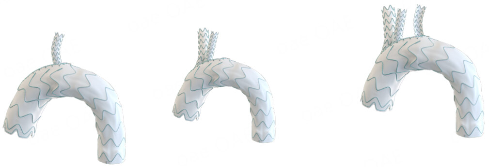
Figure 1. Left: Single-branched RELAY™ endoprosthesis. Middle: Double-branched RELAY™ endoprosthesis. Right: Triple-branched RELAY™ endoprosthesis. Figure reused from Terumo Aortic website.
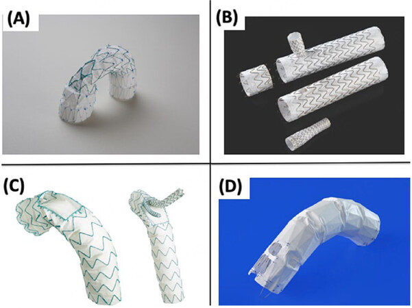
Figure 2. Examples of commercially available TER endoprostheses. (A) Zenith A-branch, (B) TAG Thoracic Branch Endoprosthesis, (C) Terumo Aortic RELAY Plus Double Branched device, and (D) Najuta. Reproduced from Fujimura et al.[46], no copyright permission is required (STM signatory).
Careful evaluation of the landing zone is imperative. In cases of dissection, it is measured using the distance from the coronary ostia and the sinotubular junction to the proximal entry of the dissection[28]. The ascending aorta possesses high velocity, consequent shear stress, and four-dimensional rotational and pulsatile movements during the cardiac cycle. Thus, the proximal and deep implantation of the device into zone 0 of the arch exposes the endoprosthesis to maximal hemodynamic pressure, which in turn increases the risk of malorientation as a result of the windsock effect[29]. Therefore, shorter and wider dimensions of the endoprosthesis are favoured, and 15% oversizing of the endoprosthesis relative to the native aortic diameter is recommended to improve FL depressurization and aneurysmal regression[30].
The triple-branched RELAY™ endoprosthesis allows for cannulation of the three-supra aortic vessels and is most suitable for long-term patency. Maintaining the left subclavian artery (LSA) patency after TER is paramount to circumvent the risk of left arm ischemia as well as SCI. Further, it avoids the subclavian steal syndrome and vertebrobasilar insufficiency, thus, leading to lower rates of stroke[31]. In cases where LSA cannulation is not possible, a single- or double-branched RELAY™ system is used, and prophylactic revascularization of the LSA (e.g., subclavian-carotid bypass) prior to TEVAR can be performed[32]. A study by Bradshaw et al. reported a 1.9% stroke rate in patients who underwent revascularization of the LSA[31]. In contrast, a stroke rate of 14.3% was reported in those who underwent TEVAR with total LSA occlusion.
Fenestrated TEVAR is another approach that has been established in treating arch pathologies while maintaining supra-aortic vessel perfusion. The Japanese Najuta™ pre-curved fenestrated stent graft is an example of fenestrated TEVAR used in TER. The Najuta™ system does not have bridging stents to fixate the fenestrations at the target-vessel ostia[33]. Another fenestrated arch endograft, the Zenith™ device from Cook Medical, uses a preloaded wire system combining a fenestration and a scallop with a covered bridging stent to fixate the fenestration to the left common carotid artery or LSA as the target vessel[34]. The distance from the femoral vessel and the curvature of the arch make the rotation of the fenestrated devices more challenging. Thus, the precision of placement requires meticulous preoperative planning and technical skills. Therefore, the arch branched devices remain more suitable for arch diseases, especially in cases of extended or complex disease[34].
Criteria for total endovascular arch repair
Despite the great potential shown by TER, OSR remains the mainstay for the treatment of aortic arch pathologies until now. Still, there are groups of patients with certain demographics and disease characteristics who are not suitable candidates for OSR. Those high-risk patients may benefit from an alternative endovascular approach that circumvents the hazards that come with CPB and HCA[35]. The decision of whether a patient is eligible or not for OSR is usually made after multidisciplinary evaluation on an individualized basis. Some high-risk features are well-established in the literature, such as older age
Besides, the use of risk prediction tools such as the American Society of Anesthesiologists (ASA) Classification and the EuroSCORE has been employed in inferring patients’ risk and determining eligibility for OSR in patients with arch disease[30,38]. Spear et al. used a multidisciplinary evaluation and an ASA III/IV to deem patients unfit for surgery[38]. In their report, they further specified the eligibility criteria to include a negative cardiac stress test, no Class III/IV heart failure, no stroke or myocardial infarction in the last year, no significant carotid stenosis, and GFR 45 mL/min/1.73 m2[38]. All these factors and more are considered in the risk stratification of patients with aortic arch disease. However, the breadth of aortic arch pathologies and associated comorbidities makes it hard to certainly stratify patients with high risks. Thus, a multidisciplinary evaluation remains the best approach to determining eligibility.
In patients with high-risk profiles for OSR, a set of morphological and disease features can determine patient eligibility for TER. Initially, for stable implantation of the endograft, an adequate landing zone is pre-determined based on parameters from preoperative imaging. These parameters include (i) a sealing zone within the ascending aorta (zone 0) less than 38 mm in diameter; and (ii) a sealing zone of at least
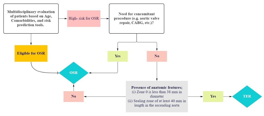
Figure 3. Flowchart (original) illustrating criteria for patients’ eligibility for TER. OSR: Open surgical repair; CABG: coronary artery bypass grafting; TER: total endoarch repair.
Anatomical requirements for TER as reported by Czerny et al.[30]
| Anatomical requirements | N |
| Ascending aorta landing zone diameter (mm) | 29-43 |
| Distal landing zone diameter (mm) | 19-43 |
| BCT and LCCA diameter (mm) | 7-20 |
| ST junction to BCT length (mm) | > 65 or > 85 |
| Distal landing zone length (mm) | 25-30 |
| BCT landing zone length (mm) | 25 |
| LCCA landing zone length (mm) | 30 |
| Proximal BCT to distal LCCA (mm) | < 45 |
As is the case with any procedure, TEVAR is associated with a significant learning curve that is reflected in the flow of the procedure as well as patient outcomes. This is evident in the retrospective single-centre study of TEVAR by Tan et al., which showed that there was a learning curve involved reflected by higher than average mean operative time, average radiation dose and mean contrast volume used during their initial experience[39].
Clinical outcomes
An appraisal of endovascular repair for aortic arch pathology requires analysis of the clinical outcomes associated therewith. Despite the minimally-invasive nature of TER providing an obvious advantage over OSR in certain cases, TER remains associated with comparable mortality rates and key complications such as technical failure, neurological injury, need for reintervention, and loss of or failure to achieve target vessel patency (TVP)[40]. These are standard metrics used to gauge the efficacy of surgical intervention on the aortic arch, and represent key challenges to the widespread adoption of TER as the gold-standard intervention for aortic arch pathologies in specific patient groups.
Technical success and target vessel patency
In the context of TER for aortic arch pathology, technical success can be defined as successful endovascular stabilisation of the aortic arch and the subsequent maintenance of aortic arch (as well as arch vessel) patency during follow-up[40]. Singh et al., in their evaluation of the RELAY™ Branched endoprosthesis in 148 patients undergoing TER between January 2019 and January 2022, reported a 99.3% (n = 147) success rate[40]. TVP was achieved in all patients and maintained during the initial 30 days postoperative. After 24 months of follow-up, an overall of 118 patients (80.2%) exhibited TVP. This included 80 (74%) patients in the double-branched group and 16 (93.7%) patients in the single-branched group who exhibited TVP at 24 months postoperatively. 100% (n = 23) of patients in the triple-branched group maintained TVP during follow-up[40]. Alsafi et al. also report a 100% technical success rate in their smaller study of 21 patients undergoing TER with RELAY™ Branched[41].
Azuma and colleagues report a 99.2% technical success rate in their cohort of 393 patients undergoing TER with the fenestrated Kawasumi Najuta™ endograft[42]. Sato et al. similarly report technical success in 97.3% of patients treated with the Najuta™ device[43]. The proximal landing zone was in Zone 0 for 86.1% of patients, while Zone 1 was selected in 13.9% of patients[43]. Iwakoshi et al. report a 91% success rate in their series of 32 patients undergoing TER with Najuta™[44]. Spear et al. report a very promising 100% technical success rate in their series of 27 patients treated with the Cook Zenith™ endoprosthesis[38]. The nonrandomized, single-arm prospective study of 9 patients who underwent TER using the Valiant™ endoprosthesis by Roselli et al. reported a 100% technical success rate[45].
Fujimura et al. highlight that TER with GORE TAG™ Thoracic Branched Endoprosthesis was anatomically feasible in 40.8% (n = 87) of patients, while TER with Relay™, Najuta™, and Zenith™ was only anatomically feasible in 24.9%, 13.6%, and 5.2% of cases respectively[46]. They suggest that TER was only feasible in 5% to 41% of patients in their series of 213 patients with arch aneurysms using fenestrated or branched endoprostheses[46]. A summary of the findings in the above subsection can be found in Table 3.
Summary of the technical success and target vessel patency subsection findings
| Study | Device | Cohort size | Technical success rate (%) |
| Singh et al.[40] | RELAY™ | 148 | 99.3 |
| Alsafi et al.[41] | RELAY™ | 21 | 100 |
| RESTORE I[47] | RELAY™ | 307 | 97.7 |
| RESTORE II[48] | RELAY™ | 173 | 97.1 |
| Azuma et al.[42] | Najuta™ | 393 | 99.2 |
| Sato et al.[43] | Najuta™ | 37 | 97.3 |
| Iwakoshi et al.[44] | Najuta™ | 32 | 91 |
| Spear et al.[38] | Cook Zenith™ | 27 | 100 |
| Roselli et al.[45] | Valiant™ | 9 | 100 |
Mortality
Having traded median sternotomy for peripheral arterial cannulation, and hypothermic circulatory arrest and cardiopulmonary bypass for simple general anaesthesia, TER is associated with improved early and long-term mortality rates relative to OSR[40,41].
Singh et al. report a 30-day mortality rate of 2.7% (n = 4) but 0 mortalities during the remainder of their follow-up period[40]. In contrast, Alsafi et al. report a 9.5% (n = 2) in-hospital mortality rate in their cohort of 21[41]. The RESTORE I and II trials were published earlier, reporting on different configurations of the RELAY™ device indicated for various aortic pathologies. The RESTORE I trial reported an in-hospital mortality of 7.2% (n = 19). This rate went lower in the RESTOE II trial featuring in-hospital mortality of 4% (n = 7)[47,48]. Additionally, Azuma et al. revealed a 1.5% (n = 6) 30-day mortality rate associated with Najuta™, while Sato et al. reported a 0% in-hospital mortality rate[42,43]. However, four deaths occurred during follow-up due to malignancy and retrograde type A aortic dissection[43]. Spear et al. report a promising 0% mortality rate associated with Zenith™, but a 3.7% 1-year aortic-related mortality[38]. O’Callaghan et al. highlight a 7% (n = 1) in-hospital mortality in patients who underwent TER with custom Cook Zenith™ and 18% (n = 3) in patients who received the non-custom endoprosthesis for proximal thoracic aneurysms[49]. Interestingly, Roselli et al. found no mortalities using the Valiant™ endoprosthesis[45]. A summary of the findings in the above subsection can be found in Table 4.
Summary of the mortality subsection findings
| Study | Device | Cohort size | Early mortality | Mean follow-up period and mortality rate | |
| Follow-up | Overall Mortality | ||||
| Singh et al.[40] | RELAY™ | 148 | 2.7% (n = 4) | 2 years | 2.7% (n = 4) |
| Alsafi et al.[41] | RELAY™ | 21 | 9.5% (n = 2) | 36 (3-183) weeks | 19% (n = 4) |
| RESTORE I[47] | RELAY™ | 307 | 7.2% (n = 19) | - | |
| RESTORE II[48] | RELAY™ | 173 | 4.0% (n = 7) | 2 years | 6.4% (n = 11) |
| Azuma et al.[42] | Najuta™ | 393 | 1.5% (n = 6) | - | |
| Sato et al.[43] | Najuta™ | 37 | 0% | 2.9 ± 2.9 years | 11.1% (n = 4) |
| Spear et al.[38] | Cook Zenith™ | 27 | 0% | 1 year | 3.7% (n = 1) |
| O’Callaghan et al.[49] | Cook Zenith™ Custom | 15 | 7% (n = 1) | - | |
| Cook Zenith™ Non-Custom | 18 | 18% (n = 3) | |||
Neurological injury
Neurological injury in the setting of aortic arch repair results primarily from ischaemia. The aetiology of cerebral injury in the context of endovascular arch repair is usually embolisation of endoluminal particulate matter (e.g., atheromatous plaque) during endovascular manipulation, or inadvertent occlusion of the carotid arteries by endovascular instrumentation[45,50]. Furthermore, spinal cord injury often occurs secondary to intercostal artery coverage by the endograft leading to compromised perfusion[51]. There is also a risk of vertebrobasilar insufficiency in patients that undergo total endovascular arch repair where the left subclavian artery is occluded and not revascularized[52].
Alsafi et al. report a 14% (n = 3) and 5% (n = 1) incidence of stroke and paraplegia following implantation of Relay™ Branched for TER[41]. Tan et al. also used Relay™ Branched in their study of 148 patients[53], which yielded a 4.1% (n = 6) stroke rate. The RESTORE I trial found that the incidence of stroke and paraplegia was 1.6% (n = 5) and 2% (n = 6), respectively, while this was 0.6% and 2.9% in RESTORE II. Azuma et al. noted that 1.8% (n = 7) of patients undergoing TER with Najuta™ suffered postoperative stroke and 0.8%
Summary of the neurological injury subsection findings
| Study | Device | Cohort size | Stroke (%) | Paraplegia (%) | SCI (%) |
| Alsafi et al.[41] | RELAY™ | 21 | 14 | 5 | - |
| Tan et al.[54] | RELAY™ | 148 | 4.1 | - | - |
| RESTORE I[47] | RELAY™ | 307 | 1.6 | 2 | - |
| RESTORE II[48] | RELAY™ | 173 | 0.6 | 2.9 | - |
| Azuma et al.[42] | Najuta™ | 393 | 1.8 | 0.8 | - |
| Sato et al.[43] | Najuta™ | 37 | 16.7 | 2.8 | - |
| Iwakoshi et al.[44] | Najuta™ | 32 | 3.1 | - | 3.1 |
| O’Callaghan et al.[49] | Cook Zenith™ | 33 | 6 | - | 6 |
| Spear et al.[38] | Cook Zenith™ | 27 | 7.4 | - | 7.4 |
| Roselli et al.[45] | Valient™ | 9 | 0 | - | - |
Reintervention
The need for reintervention is a well-known aspect of endovascular aortic repair, especially in comparison to OSR. Reintervention is typically indicated in cases involving postoperative retrograde dissection, endoleak, or endograft migration[40]. Type 1a endoleak, in particular, is suggestive of suboptimal proximal or distal sealing, or graft migration, and is therefore a familiar complication in the context of endovascular arch repair[40].
None of the patients in Singh et al. who underwent TER using single- or triple-branched RELAY™ required reintervention, while 24 (16.2%) patients that received the double-branched RELAY™ did require this post-TER[40]. Alsafi et al. reported a 10% (n = 2) reintervention rate in their series due to type 2 endoleak using RELAY™[41]. Two patients enrolled in the RESTORE I trial required surgical conversion postoperatively, while in RESTORE II, the rates of early and late reintervention were 3.5% and 7.5%, respectively. Furthermore, Azuma et al. found a 0.8% (n = 3) rate of retrograde dissection requiring reintervention associated with Najuta™[42]. Using the same device, Sato et al. reported an 8.3% (n = 3) reintervention rate during the 2.9 ± 2.9 year follow-up period[43]. This value was even higher in Iwashoki et al. at 12.5%
Summary of the reintervention subsection findings
| Study | Device | Cohort size | Reintervention (%) |
| Singh et al.[40] | Single- or -triple-branched RELAY™ | 40 | 0 |
| Double-branched RELAY™ | 108 | 16.2 | |
| Alsafi et al.[41] | RELAY™ | 21 | 10 |
| RESTORE[47] | RELAY™ | 307 | 0.7 |
| RESTORE II[48] | RELAY™ | 173 | Early: 3.5 |
| Late: 7.5 | |||
| Azuma et al.[42] | Najuta™ | 393 | 0.8 |
| Sato et al.[43] | Najuta™ | 37 | 8.3 |
| Iwakoshi et al.[44] | Najuta™ | 32 | 12.5 |
| Spear et al.[38] | Cook Zenith™ | 27 | 7.4 |
| O’Callaghan et al.[49] | Cook Zenith™ | 33 | 30.3 |
| Roselli et al.[45] | Valiant™ | 9 | 0 |
CONCLUSION
Endoarch repair using TEVAR represents the future of aortic arch repair. While FET is associated with excellent clinical outcomes, TER has achieved highly comparable results due to its novelty. Nevertheless, further studies with larger cohorts and longer follow-up periods are necessary to solidify the evidence on TER. In addition, studies directly comparing arch OSR to TER are warranted to determine superiority.
DECLARATIONS
Authors’ contributionsDevised the manuscript topic and supervised the literature search and writing process: Bashir M, Mohammed I
Performed the literature search and wrote the manuscript: Jubouri M, Al-Tawil M, Tan SZCP, Geragotellis A, Hussain M
Provided feedback on the manuscript draft: Mohammed I, Velayudhan B, Bashir M
Edited and formatted the final version of the manuscript: Jubouri M, Al-Tawil M
All authors approved this final version of the manuscript.
Availability of data and materialsData and materials available publicly in search engines/electronic databases such as PubMed, Google Scholar, EMBASE and Scopus.
Financial support and sponsorshipNone.
Conflicts of interestAll authors declared that there are no conflicts of interest.
Ethical approval and consent to participateNot applicable.
Consent for publicationNot applicable.
Copyright© The Author(s) 2023.
REFERENCES
1. Di Eusanio M, Berretta P, Cefarelli M, et al. Long-term outcomes after aortic arch surgery: results of a study involving 623 patients. Eur J Cardiothorac Surg 2015;48:483-90.
2. Tsilimparis N, Ivancev K, Kolbel T. Endovascular aortic arch repair - endovascular today. 2015. Available from: https://evtoday.com/articles/2015-nov-supplement/endovascular-aortic-arch-repair [Last accessed on 10 March 2022].
3. Antoniou GA, Mireskandari M, Bicknell CD, et al. Hybrid repair of the aortic arch in patients with extensive aortic disease. Eur J Vasc Endovasc Surg 2010;40:715-21.
4. Chiesa R, Melissano G, Tshomba Y, et al. Ten years of endovascular aortic arch repair. J Endovasc Ther 2010;17:1-11.
5. Preventza O, Garcia A, Cooley DA, et al. Total aortic arch replacement: a comparative study of zone 0 hybrid arch exclusion
6. Shrestha M, Martens A, Krüger H, et al. Total aortic arch replacement with the elephant trunk technique: single-centre 30-year results. Eur J Cardiothorac Surg 2014;45:289-95; discussion 295.
7. Shrestha M, Beckmann E, Krueger H, et al. The elephant trunk is freezing: the hannover experience. J Thorac Cardiovasc Surg 2015;149:1286-93.
8. Di Eusanio M, Pantaleo A, Murana G, et al. Frozen elephant trunk surgery-the Bologna’s experience. Ann Cardiothorac Surg 2013;2:597-605.
9. LeMaire SA, Carter SA, Coselli JS. The elephant trunk technique for staged repair of complex aneurysms of the entire thoracic aorta. Ann Thorac Surg 2006;81:1561-9; discussion 1569.
10. Svensson LG, Kim KH, Blackstone EH, et al. Elephant trunk procedure: newer indications and uses. Ann Thorac Surg 2004;78:109-16; discussion 109.
11. Safi HJ, Miller CC 3rd, Estrera AL, et al. Optimization of aortic arch replacement: two-stage approach. Ann Thorac Surg 2007;83:S815-8; discussion S824.
12. Schepens MA, Dossche KM, Morshuis WJ, van den Barselaar PJ, Heijmen RH, Vermeulen FE. The elephant trunk technique: operative results in 100 consecutive patients. Eur J Cardiothorac Surg 2002;21:276-81.
13. Jakob H, Tsagakis K, Tossios P, et al. Combining classic surgery with descending stent grafting for acute DeBakey type I dissection. Ann Thorac Surg 2008;86:95-101.
14. Di Bartolomeo R, Pantaleo A, Berretta P, et al. Frozen elephant trunk surgery in acute aortic dissection. J Thorac Cardiovasc Surg 2015;149:S105-9.
15. Geragotellis A, Surkhi AO, Jubouri M, et al. Endovascular reintervention after frozen elephant trunk: where is the evidence? J Cardiovasc Surg 2022;63:425-33.
16. Preventza O, Liao JL, Olive JK, et al. Neurologic complications after the frozen elephant trunk procedure: a meta-analysis of more than 3000 patients. J Thorac Cardiovasc Surg 2020;160:20-33.e4.
17. Di Bartolomeo R, Murana G, Di Marco L, et al. Frozen
18. Mousavizadeh M, Bashir M, Jubouri M, et al. Zone proximalization in frozen elephant trunk: what is the optimal zone for open intervention? J Cardiovasc Surg 2022;63:265-74.
19. Fiorentino M, de Beaufort HWL, Sonker U, Heijmen RH. Thoraflex hybrid as frozen elephant trunk in chronic, residual type A and chronic type B aortic dissection. Interact Cardiovasc Thorac Surg 2021;32:566-72.
20. Leone A, Beckmann E, Martens A, et al. Total aortic arch replacement with frozen elephant trunk technique: Results from two European institutes. J Thorac Cardiovasc Surg 2020;159:1201-11.
21. Ho JYK, Chow SCY, Kwok MWT, Fujikawa T, Wong RHL. Total aortic arch replacement and frozen elephant trunk. Semin Thorac Cardiovasc Surg 2021;33:656-62.
22. Jubouri M, Kayali F, Saha P, et al. Incidence of distal stent graft induced new entry
23. Kayali F, Jubouri M, Tan SZ, Mohammed I, Bashir M. Aortic remodeling in aortic dissection after frozen elephant trunk: overcoming the challenges. J Cardiovasc Surg 2022;63:434-8.
24. Kayali F, Qutaishat S, Jubouri M, Chikhal R, Tan SZCP, Bashir M. Kinking of frozen elephant trunk hybrid prostheses: incidence, mechanism, and management. Front Cardiovasc Med 2022;9:912071.
25. Bavaria JE, Appoo JJ, Makaroun MS, et al. Endovascular stent grafting
26. Makaroun MS, Dillavou ED, Kee ST, et al. Endovascular treatment of thoracic aortic aneurysms: results of the phase II multicenter trial of the GORE TAG thoracic endoprosthesis. J Vasc Surg 2005;41:1-9.
27. Kölbel T, Rostock T, Larena-Avellaneda A, Treede H, Franzen O, Debus ES. An externalized transseptal guidewire technique to facilitate guidewire stabilization and stent-graft passage in the aortic arch. J Endovasc Ther 2010;17:744-9.
28. Nordon IM, Hinchliffe RJ, Morgan R, Loftus IM, Jahangiri M, Thompson MM. Progress in endovascular management of type A dissection. Eur J Vasc Endovasc Surg 2012;44:406-10.
29. Nienaber CA, Kische S, Rehders TC, et al. Rapid pacing for better placing: comparison of techniques for precise deployment of endografts in the thoracic aorta. J Endovasc Ther 2007;14:506-12.
30. Czerny M, Rylski B, Morlock J, et al. Orthotopic branched endovascular aortic arch repair in patients who cannot undergo classical surgery. Eur J Cardiothorac Surg 2018;53:1007-12.
31. Bradshaw RJ, Ahanchi SS, Powell O, et al. Left subclavian artery revascularization in zone 2 thoracic endovascular aortic repair is associated with lower stroke risk across all aortic diseases. J Vasc Surg 2017;65:1270-9.
32. Yuan X, Mitsis A, Mozalbat D, Nienaber CA. Alternative management of proximal aortic dissection: concept and application. Indian J Thorac Cardiovasc Surg 2022;38:183-92.
33. Yokoi Y, Azuma T, Yamazaki K. Advantage of a precurved fenestrated endograft for aortic arch disease: simplified arch aneurysm treatment in Japan 2010 and 2011. J Thorac Cardiovasc Surg 2013;145:S103-9.
34. Tsilimparis N, Debus ES, von Kodolitsch Y, et al. Branched
35. Hagan PG, Nienaber CA, Isselbacher EM, et al. The international registry of acute aortic dissection (IRAD): new insights into an old disease. JAMA 2000;283:897-903.
36. Preventza O, Price MD, Amarasekara HS, et al. In the endovascular era, is elective open aortic arch surgery in elderly patients still justified? J Thorac Cardiovasc Surg 2019;158:973-9.
37. Iafrancesco M, Ranasinghe AM, Dronavalli V, et al. Open aortic arch replacement in high-risk patients: the gold standard. Eur J Cardiothorac Surg 2016;49:646-51; discussion 651.
38. Spear R, Haulon S, Ohki T, et al. Editor’s choice - subsequent results for arch aneurysm repair with inner branched endografts. Eur J Vasc Endovasc Surg 2016;51:380-5.
39. Tan GWL, Quek L, Tan BP, Pua U. Early experience and lessons learnt with customized fenestrated thoracic endovascular aortic reconstruction for aortic arch pathology in an asian population. Cardiovasc Intervent Radiol 2018;41:544-53.
40. Singh S, Surkhi AO, Tan SZCP, et al. RELAY (TM) branched-international results of vessel patency and reintervention. Front Cardiovasc Med 2022;9:962884.
41. Alsafi A, Bicknell CD, Rudarakanchana N, et al. Endovascular treatment of thoracic aortic aneurysms with a short proximal landing zone using scalloped endografts. J Vasc Surg 2014;60:1499-506.
42. Azuma T, Yokoi Y, Yamazaki K. The next generation of fenestrated endografts: results of a clinical trial to support an expanded indication for aortic arch aneurysm treatment. Eur J Cardiothorac Surg 2013;44:e156-63; discussion e163.
43. Sato H, Fukada J, Tamiya Y, et al. Long-term clinical outcomes of thoracic endovascular aortic repair for arch aneurysms with the najuta thoracic stent-graft system. Ann Vasc Dis 2020;13:384-9.
44. Iwakoshi S, Ichihashi S, Itoh H, et al. Clinical outcomes of thoracic endovascular aneurysm repair using commercially available fenestrated stent graft (Najuta endograft). J Vasc Surg 2015;62:1473-8.
45. Roselli EE, Arko FR 3rd, Thompson MM, et al. Results of the valiant mona LSA early feasibility study for descending thoracic aneurysms. J Vasc Surg 2015;62:1465-71.e3.
46. Fujimura N, Ichihashi S, Motoki M, et al. Anatomical analysis and feasibility study of next-generation fenestrated or branched stent-grafts for the treatment of arch aneurysms. J Endovasc Ther 2020;27:777-84.
47. Riambau V, Zipfel B, Coppi G, et al. Final operative and midterm results of the European experience in the RELAY Endovascular Registry for Thoracic Disease (RESTORE) study. J Vasc Surg 2011;53:565-73.
48. Zipfel B, Zaefferer P, Riambau V, et al. Worldwide results from the RESTORE II on elective endografting of thoracic aneurysms and dissections. J Vasc Surg 2016;63:1466-75.
49. O’Callaghan A, Mastracci TM, Greenberg RK, Eagleton MJ, Bena J, Kuramochi Y. Outcomes for supra-aortic branch vessel stenting in the treatment of thoracic aortic disease. J Vasc Surg 2014;60:914-20.
50. Gutsche JT, Cheung AT, McGarvey ML, et al. Risk factors for perioperative stroke after thoracic endovascular aortic repair. Ann Thorac Surg 2007;84:1195-200; discussion 1200.
51. Jiang SM, Ali Hassan SM, Nguyen G, Bisleri G. Zone 0 frozen elephant trunk for type A retrograde acute aortic dissection following endovascular stenting of the arch. J Card Surg 2021;36:2124-6.
Cite This Article
Export citation file: BibTeX | RIS
OAE Style
Jubouri M, Al-Tawil M, Tan SZCP, Geragotellis A, Hussain M, Bashir M, Velayudhan B, Mohammed I. Total endovascular aortic arch repair: is it for everyone and where is its evidence?. Vessel Plus 2023;7:5. http://dx.doi.org/10.20517/2574-1209.2022.49
AMA Style
Jubouri M, Al-Tawil M, Tan SZCP, Geragotellis A, Hussain M, Bashir M, Velayudhan B, Mohammed I. Total endovascular aortic arch repair: is it for everyone and where is its evidence?. Vessel Plus. 2023; 7: 5. http://dx.doi.org/10.20517/2574-1209.2022.49
Chicago/Turabian Style
Jubouri, Matti, Mohammed Al-Tawil, Sven Z. C. P. Tan, Alexander Geragotellis, Mariam Hussain, Mohamad Bashir, Bashi Velayudhan, Idhrees Mohammed. 2023. "Total endovascular aortic arch repair: is it for everyone and where is its evidence?" Vessel Plus. 7: 5. http://dx.doi.org/10.20517/2574-1209.2022.49
ACS Style
Jubouri, M.; Al-Tawil M.; Tan SZCP.; Geragotellis A.; Hussain M.; Bashir M.; Velayudhan B.; Mohammed I. Total endovascular aortic arch repair: is it for everyone and where is its evidence?. Vessel Plus. 2023, 7, 5. http://dx.doi.org/10.20517/2574-1209.2022.49
About This Article
Special Issue
Copyright

Data & Comments
Data
0

 Cite This Article 11 clicks

 Like This Article 8
likes
Like This Article 8
likes


Comments
Comments must be written in English. Spam, offensive content, impersonation, and private information will not be permitted. If any comment is reported and identified as inappropriate content by OAE staff, the comment will be removed without notice. If you have any queries or need any help, please contact us at support@oaepublish.com.