
Cardiac amyloidosis: the pathologist's point of view
 0
0Abstract
Cardiac amyloidosis is a well-known entity recently recognized as a common etiology of heart failure. This infiltrative disease is caused by the deposition of misfolded proteins within the heart. The most common types of cardiac amyloidosis result from fibrils composed of monoclonal immunoglobulin light chains or transthyretin. Clinical presentation is usually elusive, and this can result in diagnostic delay. Diagnosis can be reached with non-invasive methods, but it often requires tissue sampling with pathological analysis. It is fundamental to determine the type of protein being deposited in order to indicate the specific treatment. In this article, we review the main features of cardiac amyloidosis with a focus on different pathological presentations of this rare disorder.
Keywords
INTRODUCTION
Amyloidosis is a disorder caused by abnormal conformation and metabolism of proteins resulting in extracellular deposition of fibrillar material. These aggregates can alter tissue architecture and subsequently affect organ function throughout the body. The disease is usually classified according to the precursor proteins that undergo the misfolding and accumulation: more than 30 different human proteins have been recognized as precursors for the buildup of amyloid deposits. Nevertheless, only nine misfolded proteins can accumulate in the myocardium and be responsible for cardiac amyloidosis (CA)[1]. Once considered a rare disease, CA is now acknowledged as an underrecognized cause of heart failure, with a variable prevalence of up to 32% in autopsy series in older populations[2]. Since cardiac involvement is still the leading cause of morbidity and mortality in amyloidosis, early diagnosis remains critical for adequate therapeutical planning closely related to a better prognosis.
The aim of our paper is to review the available pathology literature on the gross and histological findings of cardiac involvement in amyloidosis, which are of potential value in interpreting laboratory and imaging data.
HISTORICAL NOTES
The term “amyloid” comes from the Latin amylum and the Greek amylon, meaning starch. This term was introduced in the medical field by Rudolf Virchow in 1854 to describe the small round deposits in the brain that stained pale blue on treatment with iodine and violet upon the subsequent addition of sulfuric acid. He was convinced that those structures were identical to starch and he named them “corpora amylacea”[3]. The representatives of the French and British Schools instead considered amyloid to be more closely related to cellulose and they, respectively, used the name “lardaceous” (based on the bacon-like appearance of the tissue) and “waxy” (based on the homogeneity of the material)[4]. Another historical term is the “sago spleen”, in cases of splenic involvement by amyloid, which can give a nodular appearance similar to sago starch grains. Only later observations led to the exclusion of starch and cellulose from the materials composing “amyloid”. The invention of the metachromatic stains in 1875 (particularly methyl violet stain) allowed for better detection of amyloid and localizing it in the extracellular tissue. The first description of amyloid in the cardiac tissue is reportedly the one by Soyka in 1876 who used this new method[5]. A new dye originally used to stain textile fibers was then discovered and became widely used: Congo red staining[6]. This stain was developed by the German chemist Paul Böttiger in 1884 and then sold to the Agfa company, which named it “Congo” for marketing purposes, reflecting geopolitical events of that time[7]. However, only in 1922, the German chemist Bennhold discovered the capacity of Congo red to bind to amyloid, introducing it in the diagnostic process. Initially, Congo red was not used as a histological stain to obtain the diagnosis: it was administered to patients with suspected amyloidosis and its level in plasma was supposed to decrease in patients with amyloid due to systemic tissue binding. In 1959, the first observation of amyloid by electron microscopy was reported, demonstrating a fibrillar structure that was different from collagen and similar in different tissues[8]. One of the first and most successful methods to extract amyloid from tissue was described in 1962 by Pras and colleagues (the so-called water extraction method), and it enabled the identification of the β-pleated sheet configuration of amyloid proteins and the discovery of the biochemical structure of those proteins[9].
ETIOLOGY AND PATHOGENESIS
Amyloidosis is considered a rare disease with still few epidemiological studies published. Data on the epidemiology of CA are based mainly on single-center studies or population registries. Of the 36 thus far known precursor proteins, fewer than 10 can accumulate in the myocardium and cause significant cardiac disease[10] [Table 1]. The most common types of CA result from fibrils composed of monoclonal immunoglobulin light chains (AL) or transthyretin (TTR), in either its hereditary or acquired form. AL-CA is the most frequent form with a reported prevalence of 6-10 per million[11-13]. In recent times, an increase in AL amyloidosis has been observed, uncoupled with a similar rise in incidence. This was supposed to be explained by early diagnosis and a consequent improvement in overall survival[14].
Amyloid fibril proteins and their precursors in humans. Modified from[10]
| Fibril protein | Precursor protein | Acquired or hereditary | Target organs |
| AL | Immunoglobulin light chain | A, H | All organs except CNS |
| AH | Immunoglobulin heavy chain | A | All organs except CNS |
| AA | (Apo) serum amyloid A | A | All organs except CNS |
| ATTR | Transthyretin, wild-type Transthyretin, variants | A H | Heart, lung, ligaments, tenosynovium PNS, ANS, heart, eye, leptomenynges |
| Aβ2M | β2-microglobulin, wild type β2-microglobulin, variants | A H | Muscoloskeletal system ANS |
| AApoAI | Apolipoprotein A I, variants | H | Heart, liver, kidney, PNS, testis, larynx, skin |
| AApoAII | Apolipoprotein A II, variants | H | Kidney |
| AApoAIV | Apolipoprotein A IV, wild type | A | Kidney medulla and systemic |
| AApoCII | Apolipoprotein C II, variants | H | Kidney |
| AApoCIII | Apolipoprotein C III, variants | H | Kidney |
| AGel | Gelsolin, variants | H | Kidney, PNS, cornea |
| ALys | Lysozyme, variants | H | Kidney |
| ALECT2 | Leuokcyte chemotactic factor-2 | A | Kidney, primarily |
| AFib | Fibrinogen α, variants | H | Kidney, primarily |
| ACys | Cystatin C, variants | H | CNS, PNS, skin |
| ABri | ABriPP, variants | H | CNS |
| ADan | ADanPP, variants | H | CNS |
| Aβ | Aβ precursor protein, wild type and variants | A, H | CNS |
| AαSyn | α-synuclein | A | CNS |
| ATau | Tau | A | CNS |
| APrP | Prion protein, wild type and variants | A, H | CJD, fatal insomnia, GSS syndrome |
| ACal | (Pro)calcitonin | A | C-cell thyroid tumors, kidney |
| AIAPP | Islet amyloid polypeptide | A | Islets of Langerhans, insulinomas |
| AANF | Atrial natriuretic factor | A | Cardiac atria |
| APro | Prolactin | A | Pituitary prolactinomas, aging pituitary |
| AIns | Insulin | A | Iatrogenic, local injections |
| ASPC | Lung surfactant protein | A | Lung |
| ACor | Corneodesmosin | A | Cornified epithelia, hair follicles |
| AMed | Lactadherin | A | Senile aortic media |
| AKer | Kerato-epithelin | A | Cornea, hereditary |
| ALac | Lactoferrin | A | Cornea |
| AOAAP | Odontogenic ameloblast-associated protein | A | Odontogenic tumors |
| ASem1 | Semenogelin 1 | A | Vescicula seminalis |
| AEnf | Enfurvitide | A | Iatrogenic |
| ACatK | Cathepsin K | A | Tumor associated |
| AEFEMP1 | EGF-containing fibulin-like extracellular matrix protein 1 | A | Portal veins aging-associated |
AL amyloidosis
AL amyloidosis (previously known as primary amyloidosis) results from the deposition of immunoglobin light chains produced by a plasma cell dyscrasia. The clonal plasma cells in the bone marrow produce excessive quantities of immunoglobulin fragments, usually immunoglobulin light chains, leading to their accumulation and deposition. In rare cases, immunoglobulin heavy chains or both heavy and light chains are the components of the amyloid fibrils. AL amyloidosis is usually characterized by multiorgan involvement with non-specific clinical presentation: the combination of macroglossia and periorbital purpura is virtually pathognomonic, but it occurs in less than a third of cases. The average age at diagnosis is 63 years, and about 90% of patients are older than 50. Cardiac involvement is very frequent with an occurrence of about 50%-80% and kidneys are the second most involved organ (50%-60% of patients)[15,16]. Immunoglobulin light chains can cause cardiac impairment both by mechanical stress due to deposition and by direct toxicity; this could explain the fact that sometimes AL-CA is clinically more severe than other types of amyloidosis given an apparently similar degree of amyloid deposition in the heart[17-19].Since patients affected by AL amyloidosis commonly have an underlying plasma cell dyscrasia ranging from monoclonal gammopathy of uncertain significance (MGUS) to multiple myeloma, the main treatment is chemotherapy to address the plasma cell proliferation, with regimens including bortezomib, cyclophosphamide, and dexamethasone. The aim of this strategy is to rapidly reduce the production of amyloidogenic light chains to limit the progressive damage to the involved organs, considering that the resolution of amyloid deposits is virtually impossible.
TTR amyloidosis
TTR amyloidosis (ATTR) occurs when dissociated TTR monomers misfold and assemble into amyloid fibrils. TTR is a tetramer protein produced primarily in the liver that becomes amyloidogenic when it dissociates into monomers. Two types of fibrils have been recognized as responsible for ATTR: Type A consists of C-terminal ATTR fragments and full-length TTR, whereas Type B consists only of full-length TTR. Every patient contains ATTR deposits of either Type A or B fibrils[20]. Two distinct types of ATTR exist: hereditary or mutated (ATTRmt) and wild-type (ATTRwt), also referred to as senile systemic amyloidosis, age-related amyloidosis, or senile cardiac amyloidosis. Type A fibrils appear to occur in most TTR variants including ATTRwt, while Type B fibrils are seen in some ATTRmt mutations[21]. ATTRmt is a rare autosomal dominant condition caused by mutations in the TTR gene. The most common mutation in ATTRmt is Val30Met. Clinical manifestations are mainly cardiovascular, neurological, or mixed[22]. ATTRmt patients are usually younger than ATTRwt patients, with a median age at onset of 39. Phenotypic heterogeneity is wide and results from different factors: the different TTR mutations, the geographic region of the patient, and the Val30Met aggregation (endemic or nonendemic). The curative therapy for ATTRmt is ortothopic liver transplantation or combined heart-liver transplantation, which can provide a sort of surgical gene therapy in particular for amyloidotic cardiomyopathy[23]. For patients with ATTR cardiomyopathy, new promising disease-modifying therapies have been developed, with specific targets including TTR silencing, TTR stabilization, and TTR disruption. TTR protein silencers (patisiran and inotersen) target the hepatic synthesis of TTR. Tafamidis and diflunisal are TTR stabilizers with the former being the only authorized drug for the treatment of ATTR cardiomyopathy. Other agents are under investigation for a role in TTR disruption (tauroursodeoxycholic acid and monoclonal antibodies), but their benefits are uncertain[24].
CLINICAL DIAGNOSIS
CA diagnosis can be reached through invasive or non-invasive strategies, the latter available only for ATTR. The disease should be suspected when typical signs and symptoms appear, the so-called “red flags”: proteinuria (even mild), macroglossia, skin bruises, and carpal tunnel syndrome. For what concerns the heart, congestive heart failure coupled with unexplained left ventricular (LV) hypertrophy at imaging is a prominent feature. Additional signs that can suggest CA are persistent troponin elevation, disproportionally low QRS voltage at the electrocardiogram (ECG), or early conduction system disease. The presence of discrepancy between a low-voltage ECG and an increased LV wall thickness on two-dimensional echocardiography is highly suggestive of cardiac amyloidosis (particularly AL amyloidosis)[25].
Non-invasive diagnosis (without histological sampling) is possible only for cardiac ATTR when a combination of clinical and imaging findings is documented: typical echocardiographic or cardiac magnetic resonance (CMR) findings need to be present in addition to positive scintigraphy [99mTc-pyrophosphate (PYP), 99mTc-3,3-diphosphono-1,2-propanodicarboxylic acid (DPD), or 99mTc-hydroxymethylene diphosphonate (HMDP)] and negative search in those with plasma cell dyscrasia by serum-free light chain assay, serum, and urine protein electrophoresis. The importance of ruling out AL-CA is highlighted by studies that demonstrated cardiac uptake by 99mTC-DPD also in patients with AL-CA, with cardiac uptake associated with poorer cardiac function and outcomes[26,27].
BIOPSY DIAGNOSIS
Diagnosis of CA can be reached by extracardiac biopsy coupled with typical imaging features of CA by echocardiography or CMR, in the absence of an alternative cause for increased LV wall thickness[28]. A so-called “screening biopsy” of the rectum or salivary gland may be performed, but fatty tissue biopsy or aspiration is actually the easiest method to obtain a sample for amyloid diagnosis. The procedure of abdominal fat pad fine needle aspiration requires Congo red staining and is very simple and cheap. However, although the reported specificity is high, the diagnostic sensitivity varies widely among different studies[29-31]. In particular, the use of fatty tissue aspiration is highly supported in systemic AL amyloidosis but demonstrates its limitations for diagnosis of ATTR amyloidosis, particularly ATTRwt amyloidosis, and generally cannot be used to exclude amyloidosis.
However, demonstration of amyloid fibrils in myocardial tissue through endomyocardial biopsy (EMB) remains the gold standard for the diagnosis of CA[28]. The first step of CA diagnosis is the visual recognition of a homogenous, eosinophilic substance within the myocardial interstitium by hematoxylin-eosin staining. Although Congo red is commonly applied for histological analysis when clinical suspicion of CA is present, it requires the use of polarized light microscopy and a certain degree of expertise; moreover, the difficulty of detecting the diagnostic “apple green birefringence” of the dye can cause a low sensitivity. In addition to Congo red, probably the most widely used histochemical stain to detect amyloid by pathologists worldwide, other stains can be helpful in formalin-fixed paraffin-embedded (FFPE) tissues, such as thioflavin T and S, which bind with the amyloid proteins and exhibit fluorescence in dark-field microscopy[32], and a slightly more complex to realize but highly specific histochemical dye, Sulfated Alcian Blu, which stains the mucopolysaccharide matrix associated with amyloid and gives a bright green appearance to the deposits[33]. If available, confocal laser microscopy can increase sensibility and specificity of amyloid detection in Congo red- and thioflavin T-stained tissues [Figure 1][34].
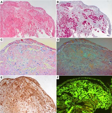
Figure 1. Endomyocardial biopsy diagnosis of cardiac amyloidosis: (A) traditional hematoxylin–eosin stain with evidence of eosinophilic interstitial deposits between cardiomyocytes; (B) Heidenhain trichrome stain highlighting amyloid in pale blue; (C,D) Congo red stain under light microscopy (C) and polarized light microscopy (D) with the typical apple-green birefringence; (E) immunohistochemical typing positive for TTR fibrils; and (F) thioflavin-T staining showing fluorescence in dark-field microscopy upon binding to amyloid fibrils. (A-F) Original magnification,× 100.
Amyloid typing
After histological confirmation, amyloid typing is essential and can be obtained with different methods: antibody-based techniques and mass spectrometry are the most widely used. Both can be applied on FFPE tissue, avoiding the need for special handling of endomyocardial biopsy samples. Antibody-based methods consist of immunofluorescence (IF), immunohistochemistry (IHC), and immunoelectron microscopy. All three techniques require a broad panel of antibodies to accurately detect the abnormal protein. In addition, each technique has its own flaws: IHC has been reported as technically complex and difficult to interpret, IF requires fresh tissue, and immune electron microscopy is expensive and requires a longer turnaround time[35]. This latter method is still used in highly specialized centers in Italy and is applied through the so-called immunogold labeling. This technique is useful for identifying biomarkers in tissues and is applicable for transmission electron microscopy and scanning electron microscopy.
Another technically complex but very specific technique is the proteomic method of the combination of laser microdissection with tandem mass spectrometry[36]. This method, first used at the Mayo Clinic in the United States, utilizes FFPE specimens as source and is highly sensitive and specific in identifying the amyloid proteins. In addition, some reports demonstrated the possibility of detecting also the underlying genetic alterations[37].
CARDIOVASCULAR INVOLVEMENT IN AMYLOIDOSIS: THE STUDY OF WHOLE HEART SPECIMENS
Amyloid can accumulate in every district of the heart in addition to the ventricular myocardium, such as atria, vessels, valves, and pericardium[35]. Whereas histological description of cardiac involvement by amyloid is well reported in the literature, comprehensive reports on whole heart specimens with detailed evaluation are rare, and their findings are summarized in Table 2[38-43]. We reviewed the literature by searching for articles (excluding case report) published in the time interval 1980-2021 with the following keywords: amyloidosis, heart, pathology and histology, autopsy, and transplantation. Only articles including analysis of whole heart specimens were considered.
Whole heart-pathology studies on cardiac amyloidosis: literature review
| Author, year ref | Source | N. cases | Mean age, years (range) | Amyloid typing | Amyloid localization | Pattern of infiltration | Valvular involvement | Vascular involvement | Amyloid quantification | Clinical correlation |
| Roberts and Waller, 1983[39] (including cases from[38]) | Autopsy | 54 | 64 (21-97) | ND | Interstitium and endocardium in all; always 1 or both atria | ND | At least 1 valve involved in all | No involvement in epicardial coronary artery Intramural vessels involved in all | ND | Hemodynamic in 16% Echo in 20% |
| Smith et al.[40], 1984 | Autopsy | 47 | Primary 57.6 (35-83) Senile 83 (70-89) | Primary vs. senile | Endocardial 70% Pericardial 36% Left atrium 91% Right atrium 81% | Nodular 49% Perifiber 28% Mixed 17% | Mitral 60% Aortic 23% Tricuspid 50% Pulmonary 30% | Present in 19 primary and 1 senile (total 43%) | ND | ND |
| Leone et al.[41], 2012 | Autopsy and HT | 9 | 56 (54-60) | 5 ATTR (unspecified) 4 AL | Computed analysis: trabecular and subendocardial layers most infiltrated | Diffuse 55.6% Segmental 22.2% Subendocardial 22.2% | ND | ND | Quantitative (%) | Echo in all CMR in 11% |
| Larsen et al.[42], 2016 | Autopsy | 108 | 75 (31-89) | 60 ATTRwt 44 AL (32 λ, 12 κ) 2 AA 1 AApoAIV 1 AL + AH | Interstitium 85% Endocardium 41% | Pericellular 66% Nodular 28% | ND | Epicardial arteries 0% Epicardial vasa vasorum 18% Epicardial veins 48% Intramyocardial arteries 72% Intramyocardial veins 9% | Semi-quantitative | ND |
| Porcari et al.[43], 2021 | Autopsy | 24 | 86 (84-91) | 12 AL 12 ATTR | Interstitium in 75% | LV non-diffuse 65% vs. diffuse 35% | ND | Vascular only in 25% | Semi-quantitative | Echo in 67% |
Gross features
The most evident macroscopic feature of CA is cardiomegaly, usually due to biventricular concentric hypertrophy. Asymmetric interventricular septal thickening can also occur[35]. The use of the term ventricular “hypertrophy” in CA is historically accepted but controversial, owing to the interstitial nature of the amyloid deposition, unrelated to myocyte enlargement[44]. The heart weight in CA can reach up to
Histological features
Ventricular myocardium
Deposition of amyloid in the myocardium can be quite variable. Infiltration has been described in terms of percentual or semiquantitative quantification, pattern, and localization. In whole hearts, the trabecular layer seems to be the most involved from the infiltration process[41]. Amyloid patterns are non-homogeneously defined in different papers, with variable descriptions of segmental, nodular, perifiber, pericellular, and diffuse deposition. Amyloid is variably associated with myocardial fibrosis, often described only as mild interstitial fibrosis but in some cases with significant extent, especially in cases with intramyocardial vascular involvement[50,51]. Cardiomyocytes adjacent to amyloid deposits can show non-specific alterations such as perinuclear halos, cytoplasmic vacuolization, and cell atrophy[52]. The presence itself of the misfolded proteins has been hypothesized as a cause of direct damage to the cardiomyocytes via apoptotic cell death, leading to cardiac dysfunction and subsequent heart failure[19,53,54].
The presence of amyloid fibrils inside the myocardium can have a direct cytotoxic effect, for both AL and TTR fragments[17,55]. This direct damage can contribute to the upregulation of an inflammatory response, positively correlating with disease severity. Histologically proven myocardial inflammation in CA has been linked with the worst prognosis in a large series of EMB, particularly in CA-AL[56]. However, different studies reported variable results in terms of association of myocardial inflammation and amyloidosis: no stress about inflammation is put in the notable autopsy series[38-42], whereas, in EMB papers, there is varied description of clusters of histiocytes, myocardial edema, and eventually plurifocal interstitial CD3+ lymphocytes in association with amyloid[51,52,57]. Isolated reports on the concomitant presence of CA and acute myocarditis are quite frequent, including in the recent context of SARS-CoV-2-associated myocardial injury[58-60].
Atria
Atrial involvement by amyloid deposits is very common. The buildup can be both intramyocardial and endocardial, the latter also being evident at gross analysis as a rugged or beadlike appearance, if very marked and diffuse[39,40]. In ATTR as well, atrial infiltration by amyloid, in particular of the left atrium, has been linked to a progressive increase in stiffness correlating with poor prognosis[61]. A different form of atrial involvement can be found in the so-called “isolated atrial amyloidosis” (IAA) caused by the accumulation of atrial natriuretic peptide or factor (ANF). IAA has typically fine deposits, unlike AL-CA and ATTR in which large plaques can occur[62,63]. This form appears to be age-related and has been commonly associated with heart failure, in which ANF levels are usually very high[64,65].
Vessels
Vascular involvement in CA is frequent and variegated. While epicardial main coronary vessels are typically spared from the condition (or limitedly involved without significant luminal obstruction), intramyocardial arteries, veins, and capillaries are usually involved in different grades by the deposits[39,42,66]. The vasa vasorum of the epicardial coronary arteries also has considerable amyloid buildups. A peculiar phenotype of intramural vessel involvement by amyloid with vascular obstruction has been widely described, sometimes linked with myocardial necrosis as well as acute onset of heart failure mimicking acute myocarditis[67-70]. This microvascular amyloid infiltration coupled with ischemic damage could explain the frequent elevation of troponin levels even in the absence of epicardial coronary lesions[71,72]. Vascular involvement is far more frequent in CA-AL compared to ATTR, acting as a relevant prognostic factor as well[40,50,73].
A different form of vascular amyloidosis is the isolated aortic amyloidosis caused by the buildup of a fibril different from light chains and TTR: the precursor is lactadherin and the accumulating fragment is called medin. Lactadherin is expressed by several kinds of cells including vascular smooth muscle cells, and the amyloid deposits are commonly located in the aortic tunica media[74-76]. This form is called AMed amyloidosis and has been demonstrated to also occur in some arterial vessels other than the aorta[77]. However, the clinical relevance is not clear yet, and no casual relation between aortic amyloid and hypertension or aneurysms has been observed.
Valves
Many works describe the histological involvement by amyloid buildups in cardiac valves, particularly in aortic valves affected by dystrophic calcification causing stenosis[78-82]. This deposition appears to be linked to the native degenerative pathology of the valve itself and the high hemodynamical stress[81]. However, only limited demonstrations of amyloid fibrils in the valvular tissue by immunohistochemistry or electron microscopy exist, and they generally exclude the presence of the common AL and TTR deposits, suggesting a different subtype for this phenomenon[83,84].
Conduction system
Typical ECG abnormalities in CA such as atrioventricular block or left anterior hemiblock prompted the detailed study of the conduction system in autopsy cases. Contrasting results were found by different authors on the localization of amyloid deposits in the specialized conduction tissue[38,67,85,86]. Most often, the sinus node, the atrioventricular node, the bundle of His, and its major branches appeared to be spared from amyloid infiltration at histological analysis. The most creditable explanation for the discordance between ECG abnormalities and amyloid sparing was given by the presence of fibrosis found in the sinoatrial and atrioventricular node, in contrast to the non-CA control group[67,86].
Pericardium
Pericardial involvement in CA is usually referred to as the presence of pericardial effusion in cases of congestive heart failure and a critical role in the prognosis[87]. However, histological demonstrations of pericardial infiltration by amyloid are scarce. When examined in the context of the autopsy series, pericardial infiltration is present in about 50% of the patients affected by CA[38,40]. In addition, a single intriguing report of isolated pericardial infiltration in AL-CA has been described but is surely an exception[88].
CONCLUSION
CA is an underestimated cause of heart failure, usually with preserved ejection fraction and restrictive hemodynamics. The role of the pathologist is not only to provide a definitive diagnosis of CA through EMB but also to help in providing precise amyloid typing since this information is essential for prognostic and therapeutic purposes.
DECLARATIONS
Authors’ contributionsDraft the manuscript: De Gaspari M
Complete and revise the paper: Finato N, Basso C
Availability of data and materialsNot applicable.
Financial support and sponsorshipMDG and CB are supported by the Registry for cardio-cerebro-vascular pathology and sudden death in the young of the Veneto Region, Italy.
Conflicts of interestAll authors declared that there are no conflicts of interest.
Ethical approval and consent to participateNot applicable.
Consent for publicationNot applicable.
Copyright© The Author(s) 2022.
REFERENCES
1. Benson MD, Buxbaum JN, Eisenberg DS, et al. Amyloid nomenclature 2018: recommendations by the International Society of Amyloidosis (ISA) nomenclature committee. Amyloid 2018;25:215-9.
2. Maurer MS, Elliott P, Comenzo R, Semigran M, Rapezzi C. Addressing common questions encountered in the diagnosis and management of cardiac amyloidosis. Circulation 2017;135:1357-77.
5. Soyka J. Über amyloide Degeneration. Prag. med. Wchnschr 165 (1876).
8. Cohen AS, Calkins E. Electron microscopic observations on a fibrous component in amyloid of diverse origins. Nature 1959;183:1202-3.
9. Pras M, Schubert M, Zucker-Franklin D, Rimon A, Franklin EC. The characterization of soluble amyloid prepared in water. J Clin Invest 1968;47:924-33.
10. Benson MD, Buxbaum JN, Eisenberg DS, et al. Amyloid nomenclature 2020: update and recommendations by the International Society of Amyloidosis (ISA) nomenclature committee. Amyloid 2020;27:217-22.
11. Kyle R, Linos A, Beard C, et al. Incidence and natural history of primary systemic amyloidosis in Olmsted County, Minnesota, 1950 through 1989. Blood 1992;79:1817-22.
12. Pinney JH, Smith CJ, Taube JB, et al. Systemic amyloidosis in England: an epidemiological study. Br J Haematol 2013;161:525-32.
13. Hemminki K, Li X, Försti A, Sundquist J, Sundquist K. Incidence and survival in non-hereditary amyloidosis in Sweden. BMC Public Health 2012;12:974.
14. Quock TP, Yan T, Chang E, Guthrie S, Broder MS. Epidemiology of AL amyloidosis: a real-world study using US claims data. Blood Adv 2018;2:1046-53.
16. Muchtar E, Dispenzieri A, Magen H, et al. Systemic amyloidosis from A (AA) to T (ATTR): a review. J Intern Med 2021;289:268-92.
17. Liao R, Jain M, Teller P, et al. Infusion of light chains from patients with cardiac amyloidosis causes diastolic dysfunction in isolated mouse hearts. Circulation 2001;104:1594-7.
18. Brenner DA, Jain M, Pimentel DR, et al. Human amyloidogenic light chains directly impair cardiomyocyte function through an increase in cellular oxidant stress. Circ Res 2004;94:1008-10.
19. Shi J, Guan J, Jiang B, et al. Amyloidogenic light chains induce cardiomyocyte contractile dysfunction and apoptosis via a non-canonical p38alpha MAPK pathway. Proc Natl Acad Sci U S A 2010;107:4188-93.
20. Bergström J, Gustavsson A, Hellman U, et al. Amyloid deposits in transthyretin-derived amyloidosis: cleaved transthyretin is associated with distinct amyloid morphology. J Pathol 2005;206:224-32.
21. Suhr OB, Lundgren E, Westermark P. One mutation, two distinct disease variants: unravelling the impact of transthyretin amyloid fibril composition. J Intern Med 2017;281:337-47.
22. Rapezzi C, Quarta CC, Obici L, et al. Disease profile and differential diagnosis of hereditary transthyretin-related amyloidosis with exclusively cardiac phenotype: an Italian perspective. Eur Heart J 2013;34:520-8.
23. Rapezzi C, Quarta CC, Riva L, et al. Transthyretin-related amyloidoses and the heart: a clinical overview. Nat Rev Cardiol 2010;7:398-408.
24. Kittleson MM, Maurer MS, Ambardekar AV, et al. American Heart Association Heart Failure and Transplantation Committee of the Council on Clinical Cardiology. Cardiac amyloidosis: evolving diagnosis and management: a scientific statement from the American heart association. Circulation 2020;142:e7-e22.
25. Murtagh B, Hammill SC, Gertz MA, Kyle RA, Tajik AJ, Grogan M. Electrocardiographic findings in primary systemic amyloidosis and biopsy-proven cardiac involvement. Am J Cardiol 2005;95:535-7.
26. Rapezzi C, Quarta CC, Guidalotti PL, et al. Usefulness and limitations of 99mTc-3,3-diphosphono-1,2-propanodicarboxylic acid scintigraphy in the aetiological diagnosis of amyloidotic cardiomyopathy. Eur J Nucl Med Mol Imaging 2011;38:470-8.
27. Quarta CC, Zheng J, Hutt D, et al. 99mTc-DPD scintigraphy in immunoglobulin light chain (AL) cardiac amyloidosis. Eur Heart J Cardiovasc Imaging 2021;22:1304-11.
28. Garcia-Pavia P, Rapezzi C, Adler Y, et al. Diagnosis and treatment of cardiac amyloidosis: a position statement of the ESC Working Group on Myocardial and Pericardial Diseases. Eur Heart J 2021;42:1554-68.
29. Gameren II, Hazenberg BP, Bijzet J, van Rijswijk MH. Diagnostic accuracy of subcutaneous abdominal fat tissue aspiration for detecting systemic amyloidosis and its utility in clinical practice. Arthritis Rheum 2006;54:2015-21.
30. Fine NM, Arruda-Olson AM, Dispenzieri A, et al. Yield of noncardiac biopsy for the diagnosis of transthyretin cardiac amyloidosis. Am J Cardiol 2014;113:1723-7.
31. Quarta CC, Gonzalez-Lopez E, Gilbertson JA, et al. Diagnostic sensitivity of abdominal fat aspiration in cardiac amyloidosis. Eur Heart J 2017;38:1905-8.
32. Picken MM, Herrera GA. Thioflavin T Stain: an easier and more sensitive method for amyloid detection. In: Picken MM, Herrera GA, Dogan A, editors. Amyloid and Related Disorders. Cham: springer International Publishing; 2015. pp. 225-7.
33. Pomerance A, Slavin G, McWatt J. Experience with the sodium sulphate-Alcian Blue stain for amyloid in cardiac pathology. J Clin Pathol 1976;29:22-6.
34. Castellani C, Fedrigo M, Frigo AC, et al. Application of confocal laser scanning microscopy for the diagnosis of amyloidosis. Virchows Arch 2017;470:455-63.
35. Maleszewski JJ. Cardiac amyloidosis: pathology, nomenclature, and typing. Cardiovasc Pathol 2015;24:343-50.
36. Shen K, Sun W, Sun J, et al. [Classification of amyloidosis by laser micro-dissection and mass spectrometry based proteomic analysis]. Zhonghua Xue Ye Xue Za Zhi 2015;36:99-102.
37. Dasari S, Theis JD, Vrana JA, et al. Clinical proteome informatics workbench detects pathogenic mutations in hereditary amyloidoses. J Proteome Res 2014;13:2352-8.
38. Buja L, Khoi NB, Roberts WC. Clinically significant cardiac amyloidosis. The American Journal of Cardiology 1970;26:394-405.
39. Roberts WC, Waller BF. Cardiac amyloidosis causing cardiac dysfunction: analysis of 54 necropsy patients. The American Journal of Cardiology 1983;52:137-46.
40. Smith TJ, Kyle RA, Lie J. Clinical Significance of Histopathologic Patterns of Cardiac Amyloidosis. Mayo Clin Proc 1984;59:547-55.
41. Leone O, Longhi S, Quarta CC, et al. New pathological insights into cardiac amyloidosis: implications for non-invasive diagnosis. Amyloid 2012;19:99-105.
42. Larsen BT, Mereuta OM, Dasari S, et al. Correlation of histomorphological pattern of cardiac amyloid deposition with amyloid type: a histological and proteomic analysis of 108 cases. Histopathology 2016;68:648-56.
43. Porcari A, Bussani R, Merlo M, et al. Incidence and characterization of concealed cardiac amyloidosis among unselected elderly patients undergoing post-mortem examination. Front Cardiovasc Med 2021;8:749523.
44. Elliott P, Andersson B, Arbustini E, et al. Classification of the cardiomyopathies: a position statement from the european society of cardiology working group on myocardial and pericardial diseases. Eur Heart J 2008;29:270-6.
46. Dinwoodey DL, Skinner M, Maron MS, Davidoff R, Ruberg FL. Light-chain amyloidosis with echocardiographic features of hypertrophic cardiomyopathy. Am J Cardiol 2008;101:674-6.
47. Philippakis AA, Falk RH. Cardiac amyloidosis mimicking hypertrophic cardiomyopathy with obstruction: treatment with disopyramide. Circulation 2012;125:1821-4.
48. Basso C, Michaud K, d'Amati G, et al. Association for European Cardiovascular Pathology. Cardiac hypertrophy at autopsy. Virchows Arch 2021;479:79-94.
49. Feng D, Edwards WD, Oh JK, et al. Intracardiac thrombosis and embolism in patients with cardiac amyloidosis. Circulation 2007;116:2420-6.
50. Neben-Wittich MA, Wittich CM, Mueller PS, Larson DR, Gertz MA, Edwards WD. Obstructive intramural coronary amyloidosis and myocardial ischemia are common in primary amyloidosis. Am J Med 2005;118:1287.
51. Pucci A, Aimo A, Musetti V, et al. Amyloid deposits and fibrosis on left ventricular endomyocardial biopsy correlate with extracellular volume in cardiac amyloidosis. J Am Heart Assoc 2021;10:e020358.
52. Frenzel H, Schwartzkopff B, Kuhn H, et al. Cardiac amyloid deposits in endomyocardial biopsies. Light microscopic, ultrastructural, and immunohistochemical studies. Am J Clin Pathol 1986;85:674-80.
53. Sikkink LA, Ramirez-Alvarado M. Cytotoxicity of amyloidogenic immunoglobulin light chains in cell culture. Cell Death Dis 2010;1:e98.
54. Mishra S, Guan J, Plovie E, et al. Human amyloidogenic light chain proteins result in cardiac dysfunction, cell death, and early mortality in zebrafish. Am J Physiol Heart Circ Physiol 2013;305:H95-103.
55. Dittloff KT, Iezzi A, Zhong JX, Mohindra P, Desai TA, Russell B. Transthyretin amyloid fibrils alter primary fibroblast structure, function, and inflammatory gene expression. Am J Physiol Heart Circ Physiol 2021;321:H149-60.
56. Siegismund CS, Escher F, Lassner D, et al. Intramyocardial inflammation predicts adverse outcome in patients with cardiac AL amyloidosis. Eur J Heart Fail 2018;20:751-7.
57. Kotecha T, Martinez-Naharro A, Treibel TA, et al. Myocardial Edema and Prognosis in Amyloidosis. J Am Coll Cardiol 2018;71:2919-31.
58. Lim HE, Pak HN, Kim YH. Acute myocarditis associated with cardiac amyloidosis manifesting as transient complete atrioventricular block and slow ventricular tachycardia. Int J Cardiol 2006;109:395-7.
59. Basso C, Leone O, Rizzo S, et al. Pathological features of COVID-19-associated myocardial injury: a multicentre cardiovascular pathology study. Eur Heart J 2020;41:3827-35.
60. Bois MC, Boire NA, Layman AJ, et al. COVID-19-Associated Nonocclusive Fibrin Microthrombi in the Heart. Circulation 2021;143:230-43.
61. Bandera F, Martone R, Chacko L, et al. Clinical importance of left atrial infiltration in cardiac transthyretin amyloidosis. JACC Cardiovasc Imaging 2022;15:17-29.
62. Lool L. Isolated atrial amyloidosis: a clinicopathologic study indicating increased prevalence in chronic heart disease. Human Pathology 1993;24:602-7.
63. Fayyaz AU, Bois MC, Dasari S, et al. Amyloidosis in surgically resected atrial appendages: a study of 345 consecutive cases with clinical implications. Mod Pathol 2020;33:764-74.
64. Kaye GC, Butler MG, d'Ardenne AJ, Edmondson SJ, Camm AJ, Slavin G. Isolated atrial amyloid contains atrial natriuretic peptide: a report of six cases. Br Heart J 1986;56:317-20.
65. Pucci A, Wharton J, Arbustini E, et al. Atrial amyloid deposits in the failing human heart display both atrial and brain natriuretic peptide-like immunoreactivity. J Pathol 1991;165:235-41.
66. Wittich CM, Neben-Wittich MA, Mueller PS, Gertz MA, Edwards WD. Deposition of amyloid proteins in the epicardial coronary arteries of 58 patients with primary systemic amyloidosis. Cardiovasc Pathol 2007;16:75-8.
67. Smith RR, Hutchins GM. Ischemic heart disease secondary to amyloidosis of intramyocardial arteries. The American Journal of Cardiology 1979;44:413-7.
68. Saffitz JE, Sazama K, Roberts WC. Amyloidosis limited to small arteries causing angina pectoris and sudden death. The American Journal of Cardiology 1983;51:1234-5.
69. Barbour DJ, Roberts WC. Frequency of acute and healed myocardial infarcts in fatal cardiac amyloidosis. The American Journal of Cardiology 1988;62:1134-5.
70. Pasotti M, Agozzino M, Concardi M, Merlini G, Rapezzi C, Arbustini E. Obstructive intramural coronary amyloidosis: a distinct phenotype of cardiac amyloidosis that can cause acute heart failure. Eur Heart J 2006;27:1810.
71. Damy T, Jaccard A, Guellich A, et al. Identification of prognostic markers in transthyretin and AL cardiac amyloidosis. Amyloid 2016;23:194-202.
72. Kristen AV, Maurer MS, Rapezzi C, Mundayat R, Suhr OB, Damy T. THAOS investigators. Impact of genotype and phenotype on cardiac biomarkers in patients with transthyretin amyloidosis - report from the Transthyretin Amyloidosis Outcome Survey (THAOS). PLoS One 2017;12:e0173086.
73. Olson LJ, Gertz MA, Edwards WD, et al. Senile cardiac amyloidosis with myocardial dysfunction. Diagnosis by endomyocardial biopsy and immunohistochemistry. N Engl J Med 1987;317:738-42.
74. Cornwell GG 3rd, Westermark P, Murdoch W, Pitkänen P. Senile aortic amyloid. A third distinctive type of age-related cardiovascular amyloid. Am J Pathol 1982;108:135-9.
75. Mucchiano G, Cornwell GG 3rd, Westermark P. Senile aortic amyloid. Evidence for two distinct forms of localized deposits. Am J Pathol 1992;140:871-7.
76. Häggqvist B, Näslund J, Sletten K, et al. Medin: an integral fragment of aortic smooth muscle cell-produced lactadherin forms the most common human amyloid. Proc Natl Acad Sci U S A 1999;96:8669-74.
77. Peng S, Glennert J, Westermark P. Medin-amyloid: a recently characterized age-associated arterial amyloid form affects mainly arteries in the upper part of the body. Amyloid 2005;12:96-102.
78. Goffin Y. Microscopic amyloid deposits in the heart valves: a common local complication of chronic damage and scarring. J Clin Pathol 1980;33:262-8.
80. Ladefoged C, Rohr N. Amyloid deposits in aortic and mitral valves. A clinicopathological investigation of material from 100 consecutive heart valve operations. Virchows Arch A Pathol Anat Histopathol 1984;404:301-12.
81. Kristen AV, Schnabel PA, Winter B, et al. High prevalence of amyloid in 150 surgically removed heart valves--a comparison of histological and clinical data reveals a correlation to atheroinflammatory conditions. Cardiovasc Pathol 2010;19:228-35.
82. Audet A, Côté N, Couture C, et al. Amyloid substance within stenotic aortic valves promotes mineralization. Histopathology 2012;61:610-9.
83. Iwata T, Nakamura H, Nagasawa T, et al. Amyloid deposits in heart valves. Acta Pathol Jpn 1982;32:23-9.
84. Goffin YA, Murdoch W, Cornwell GG 3rd, Sorenson GD. Microdeposits of amyloid in sclerocalcific heart valves: a histochemical and immunofluorescence study. J Clin Pathol 1983;36:1342-9.
85. James TN. Pathology of the cardiac conduction system in amyloidosis. Ann Intern Med 1966;65:28-36.
86. Ridolfi RL, Bulkley BH, Hutchins GM. The conduction system in cardiac amyloidosis. The American Journal of Medicine 1977;62:677-86.
87. Yuda S, Hayashi T, Yasui K, et al. Pericardial Effusion and Multiple Organ Involvement Are Independent Predictors of Mortality in Patients with Systemic Light Chain Amyloidosis. Intern Med 2015;54:1833-40.
Cite This Article
Export citation file: BibTeX | RIS
OAE Style
De Gaspari M, Finato N, Basso C. Cardiac amyloidosis: the pathologist's point of view. Vessel Plus 2022;6:57. http://dx.doi.org/10.20517/2574-1209.2022.05
AMA Style
De Gaspari M, Finato N, Basso C. Cardiac amyloidosis: the pathologist's point of view. Vessel Plus. 2022; 6: 57. http://dx.doi.org/10.20517/2574-1209.2022.05
Chicago/Turabian Style
De Gaspari, Monica, Nicoletta Finato, Cristina Basso. 2022. "Cardiac amyloidosis: the pathologist's point of view" Vessel Plus. 6: 57. http://dx.doi.org/10.20517/2574-1209.2022.05
ACS Style
De Gaspari, M.; Finato N.; Basso C. Cardiac amyloidosis: the pathologist's point of view. Vessel Plus. 2022, 6, 57. http://dx.doi.org/10.20517/2574-1209.2022.05
About This Article
Special Issue
Copyright

Data & Comments
Data
0

 Cite This Article 13 clicks

 Like This Article 1
likes
Like This Article 1
likes


Comments
Comments must be written in English. Spam, offensive content, impersonation, and private information will not be permitted. If any comment is reported and identified as inappropriate content by OAE staff, the comment will be removed without notice. If you have any queries or need any help, please contact us at support@oaepublish.com.