
Contemporary pharmacological treatment strategies for patients with angina and unobstructed coronary arteries (ANOCA) due to coronary microvascular dysfunction

Abstract
In recent years, there has been important progress in the evolving field of coronary vasomotor disorders regarding diagnostic assessments and therapeutic strategies. It is now commonly accepted that patients with angina and unobstructed coronary arteries (ANOCA) frequently suffer from coronary vasomotor disorders. The latter can be reliably diagnosed using invasive diagnostic procedures. They comprise the detection of epicardial and/or microvascular spasm, impaired coronary vasodilatation, and enhanced microvascular resistance. As these mechanisms may overlap in one given patient, various endotypes of disease can be distinguished. Although evidence from randomized clinical trials in this setting is still sparse, it has been suggested that - in addition to strict risk factor control-targeted pharmacological therapies/treatments based on the identified mechanism of disease can improve symptoms and prognosis. In patients with coronary spasm as the predominant mechanism, first-line treatment consists of high-dose calcium channel blockers and nitrates. In patients with impaired vasodilatation or enhanced microvascular resistance, beta-blockers, angiotensin-converting enzyme inhibitors, and statins represent first-line treatment. In the group of patients with symptoms refractory to first-line medication, second-line drugs such as nicorandil, molsidomine, ranolazine, ivabradine, and others for microvascular dysfunction are available. Moreover, ongoing studies in this area are evaluating the usefulness of newer pharmacological agents such as endothelin-receptor antagonists or soluble guanylate cyclase stimulators. This article summarizes the currently available evidence for pharmacological treatment strategies in patients with ANOCA due to coronary microvascular dysfunction.
Keywords
INTRODUCTION
Coronary microvascular dysfunction (CMD) is still an under-recognized cardiac condition that can be responsible for the impairment of myocardial perfusion and ischemia[1,2]. In patients who show symptoms of coronary artery disease (CAD), CMD has a high prevalence, especially in women (54%)[3]. It is associated with adverse outcomes[4-9], but currently there are no therapy guidelines available because large-scale evidence-based data are missing. In 2020, an EAPCI Expert Consensus Document on ischemia with non-obstructive coronary arteries endorsed by the “Coronary Vasomotor Disorders International Study Group” (COVADIS) was published which lists therapy recommendations[10]. Nevertheless, treatment tactics often remain empiric and should be adapted to the underlying pathomechanism as much as possible. Given the large number of patients affected by this condition, randomized studies for the evaluation of optimal treatment strategies are needed. This review focuses on pharmacotherapy in patients with angina and unobstructed coronary arteries (ANOCA) due to CMD.
Definition of CMD
Coronary microvessels with a diameter < 500 μm represent prearterioles and arterioles, which are characterized by a very large drop in pressure along their length. Arterioles are the spot of metabolic regulation of myocardial blood flow since their tone is altered by substances produced during myocardial metabolism[11]. CMD is defined as a mismatch of myocardial blood supply and oxygen consumption due to a dysregulation of coronary microvessels[1]. Both structural and functional changes in the microvasculature are possible causes. Vascular remodeling and rarefaction, perivascular fibrosis, and endothelial and vascular smooth muscle cell dysfunction have been described in CMD. The current classification of CMD contains four types: Type 1 is CMD in the absence of CAD and myocardial diseases; Type 2 is CMD in the presence of myocardial diseases; Type 3 is CMD in the presence of obstructive CAD; and Type 4 is iatrogenically induced CMD[1].
CMD in the absence of myocardial diseases and obstructive coronary artery disease
In Type 1 CMD, several disturbances have been defined that may account for the clinical presentation of angina. They comprise coronary microvascular spasm[12] and impaired coronary microvascular dilatation[13]/enhanced microvascular resistance. Initially, Type 1 CMD was regarded as the functional equivalent of traditional coronary risk factors (smoking, hypertension, hyperlipidemia, and diabetes). However, the results of the “Women’s Ischemia Syndrome Evaluation” showed that traditional cardiovascular risk factors account for less than 20% of the observed variability in coronary flow reserve in response to adenosine[14]. Therefore, other mechanisms seem to be involved, and Type 1 CMD is only partly reversible by reducing the burden of common cardiovascular risk factors.
Interventional diagnostic procedure
Patients with CMD often present with angina or dyspnea suggestive for obstructive epicardial CAD, and many of them undergo invasive coronary angiography. However, the rate of obstructive CAD in this population is under 40%[15]. Therefore, the remaining ANOCA patients have a high probability to suffer from myocardial ischemia instigated by coronary vasomotor disorders including CMD[12,16,17]. To expedite an immediate diagnosis in these patients, invasive methods to evaluate the coronary microcirculation during coronary angiography are of great significance. For this purpose, several methods have been established during the last decades. One of these methods, called interventional diagnostic procedure (IDP), includes coronary flow reserve (CFR) and coronary microvascular resistance (MVR) measurements by intracoronary Doppler or thermodilution and acetylcholine spasm provocation testing[18]. IDP allows the evaluation of decreased vasodilation of the microvasculature in reaction to adenosine. In addition, it can also show an increased microvascular hyperconstrictive reaction to acetylcholine. The primary pathophysiology of CMD includes a pathological reaction of the coronary microvessels to structural, metabolic, and neurohumoral vasoactive provocations resulting in compromised vasodilatory capacity and/or enhanced microvascular vasoconstriction. Therefore, complete invasive assessments of coronary microvascular function contain the evaluation of the vasodilator as well as the vasoconstrictor microvascular responses.
Assessment of the vasoconstrictor element is confined to invasive methods[19]. The standardized criteria for microvascular spasm focus on the patient’s reproduction of symptoms and ischemic electrocardiogram (ECG) alterations in the absence of epicardial spasm (< 90%) during the provocation test because there is currently no imaging technique available to illustrate the coronary microvascular function in vivo[20].
CFR and/or MVR are usually measured as a ratio at rest and during hyperemia induced by adenosine, regadenoson, or dipyridamole. Those agents can be administered intravenously or intracoronary[21,22]. Myocardial oxygen demand and coronary blood flow show an almost linear relationship. Hence, healthy subjects have a CFR up to 5[23]. CFR may be decreased in patients with CMD but also in patients with an epicardial stenosis. Therefore, CFR can only be regarded as a measure of microvascular function in patients with unobstructed coronary arteries as it does not discriminate microvascular and epicardial disease. CFR has been demonstrated to rely on the patient’s hemodynamics at rest[24]. That is why it was criticized for being dependent on baseline coronary flow[25]. Due to these disadvantages of CRF, parameters autonomous of baseline coronary flow have been established. Those techniques measure the minimal MVR during maximal hyperemia. For this reason, Doppler and thermodilution wires designed with a supplementary distal pressure sensor are offered. They enable real-time measurements of the distal arterial pressure (Pd) and coronary flow or transit time. Contrary to CFR, MVR can measure microvascular function in the case of epicardial stenosis.
Definition of endotypes
In the context of IDP, different endotypes of vasomotion disorders can be distinguished, which are demonstrated in Figure 1. First, pathological vasodilation and pathological vasoconstriction can be differentiated. The endotype pathological vasoconstriction can then be further subdivided into epicardial vs. microvascular. The endotype pathological vasodilation, on the other hand, can be subdivided into pathological CFR vs. pathological resistance. The following statements attempt to highlight a targeted pharmacological therapy for ANOCA patients (Type 1 CMD) that differentiates according to the endotypes identified in IDP. Those patients show no macroscopic anatomical pathologies (absence of myocardial diseases and obstructive coronary artery diseases). However, structural, anatomic changes may still be present in the microvasculature (e.g., vascular remodeling and rarefaction and perivascular fibrosis). Since IDP also captures these microvascular changes by measuring MVR, the targeted treatment approach based on the IDP results is able to address both structural/anatomical and functional abnormalities.
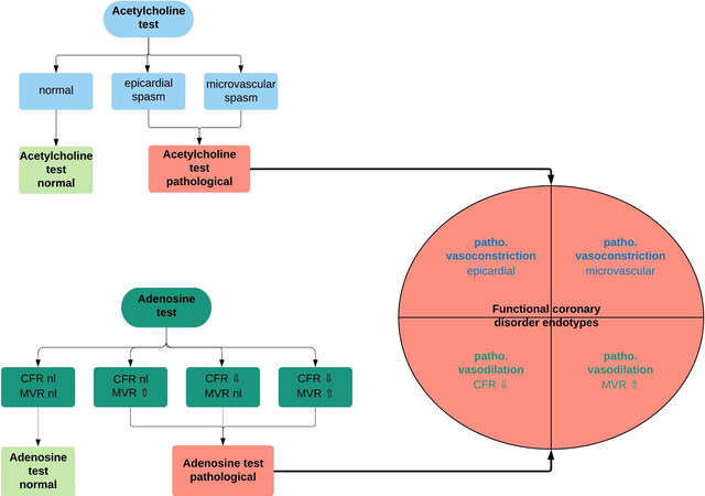
Figure 1. Definition of functional coronary disorder endotypes as determined by the interventional diagnostic procedure. Based on the consecutive testing of the coronary vasomotor response to adenosine and acetylcholine during the interventional diagnostic procedure, different functional coronary disorder endotypes can be distinguished, which can also be detected in combination. CFR: Coronary flow reserve; MVR: microvascular resistance; nl: normal; patho: pathological.
TARGETED PHARMACOLOGICAL THERAPIES
The CORonary MICrovascular Angina (CorMicA) trial[18] randomized 151 patients with non-obstructive coronary disease to a stratified medical therapy group guided by the results of intracoronary testing vs. a control group receiving standard care. The results show a significant reduction of anginal symptoms in the tailored therapy group. Before offering tailored pharmacotherapy to ANOCA patients, the cardiovascular risk profile of the patient should be evaluated. Studies have shown that common cardiovascular risk factors also affect the coronary microvasculature[26]. Therefore, a reduction of risk factors may also be beneficial for this patient population.
Predominant mechanism: coronary spasms
Calcium channel blockers
Calcium channel blockers (CCBs) prevent the voltage-dependent L-type calcium channel activation, and consequently they are smooth muscle dilators and negative inotropic and chronotropic substances. Several studies have stated the usefulness of CCBs in decreasing angina frequency in patients with epicardial vasospasms[27,28]. Treatment of microvascular spasms is less well established. CCBs are reportedly of limited efficacy in patients with microvascular angina[29,30]. Conversely, other studies have shown favorable effects of nisoldipine and diltiazem, indicating that at least a subset of patients with microvascular vasospastic angina benefits from CCB treatment[30]. Hence, CCBs are endorsed and are often effective in patients with CMD. A protective effect of CCBs is also supported in animal studies. For instance, amlodipine prevents inward remodeling in porcine coronary microvessels[31]. If patients report frequent attacks of angina pectoris and IDP reveals microvascular spasms (i.e., reproduction of angina during ACh provocation test without epicardial spasm but with ischemic ECG changes), a CCB treatment should be initiated as in the case of epicardial vasospasms. In addition, short acting nitrates are suggested to relieve spontaneous attacks of angina. However, patients often report a limited effect of these drugs. In these cases, sublingual nitrendipine for a quick relief of symptoms is recommended.
Benidipine is a potent and long-acting CCB, which inhibits not only the L- and N- but also the T-type calcium channel and controls the vasoconstriction and vasodilation of renal efferent arterioles[32]. It also inhibits aldosterone production[33,34], directly inhibits aldosterone-induced mineralocorticoid receptor activation[35,36], and has a sodium diuretic effect[37]. Benidipine was initially licensed for use in Japan and selected Southeast Asian countries and later in Turkey. A meta-analysis that compared the prognostic effects of four CCBs (amlodipine, nifedipine, diltiazem, and benidipine) showed a significant superiority of benidipine regarding the occurrence of major adverse cardiovascular events in Japanese patients with a history of vasospastic angina attacks[38]. In addition, use of benidipine showed significantly better control of angina symptoms compared with diltiazem[39]. Benidipine improves endothelial dysfunction beyond blood pressure control in patients with coronary vasospasm. Upregulation of the nitric oxide (NO) cGMP system by benidipine may somewhat add to the improvement[40]. Overall, the dihydropyridine (DHP) class may be more beneficial for vascular endothelial function than the non-dihydropyridine (non-DHP) classes of CCBs. However, since DHP CCBs can lead to reflex tachycardia, we usually use them in patients with a resting heart rate below 70 bpm. Patients with a resting heart rate above 70 bpm should be treated with a non-DHP CCB.
Nitrates
Besides CCBs, nitrates are commonly used as concomitant drugs for the treatment of epicardial vasospastic angina. Multiple studies have reported inconclusive results regarding the effectiveness of short-acting nitrates on angina pectoris symptoms in patients with CMD[41,42]. It has been assumed that nitrates have different effects in the epicardial coronary arteries compared to the microvascular coronary arteries due to different signaling pathways[43]. Studies on long-acting nitrates have usually shown no helpful effect, and they are therefore not suggested as first-line drugs in these patients[44]. However, long-term consumption of nicorandil is not associated with poor clinical outcomes, in contrast to conventional nitrates[44]. Nicorandil has a nitrate-like influence, releasing NO and causing vasodilation via cGMP signaling pathways[45,46]. However, it also acts on K+ATP channels of vascular smooth muscle cells and causes dilation of coronary microvessels and peripheral resistance arteries[47,48]. Unlike conventional nitrates, nicorandil is not linked to tolerance or rebound angina, perhaps due to its two separate mechanisms of action[47,49]. Furthermore, there is some indication that long-term use of nicorandil improves endothelial function alongside decreases in biomarkers of oxidative stress and systemic inflammation[50].
In our experiences, short-acting nitrates such as sublingual nitroglycerin spray are frequently effective in relieving chest pain symptoms in patients with vasospastic angina, particularly when given in addition to a CCB treatment. However, except for nicorandil, long-acting nitrates in the form of tablets are less effective at relieving symptoms in vasospastic angina patients.
Predominant mechanism: impaired vasodilation/enhanced microvascular resistance
Beta-blockers
In patients who show impaired vasodilation and enhanced microvascular resistance during IDP, the underlying pathophysiology is assumed to be based on microvascular remodeling (e.g., arteriolar narrowing, inward remodeling, or capillary rarefaction)[51]. A principal goal of pharmacotherapy for the coronary microcirculation is a “normalization” of transformed microvascular structure (“re-remodeling”). Conventional beta-blockers appear to have an inadequate effect on re-remodeling. However, they act by blocking catecholamine-induced increase in heart rate, blood pressure, and myocardial contractility, thereby reducing myocardial oxygen demand and consumption[52]. This is reflected in a reduced resting coronary flow causing an increase in coronary blood flow reserve[53]. A class of beta-blockers that releases NO seems especially beneficial in cases of a predominant vasodilation impairment with additional coronary spasms diagnosed during acetylcholine provocation testing[54,55].
Angiotensin-converting enzyme inhibitors/Angiotensin II receptor blockers
Endothelium-dependent vasodilation can at least partly be restored by several medications. These include angiotensin-converting enzyme inhibitors (ACE-I) or angiotensin II receptor blockers (ARB)[56-58]. ACE-I and ARB decrease production of angiotensin II, which has vasoconstrictive properties. They also decrease degradation of endothelial bradykinin, which stimulates production of NO and other vasodilators. ACE-I and ARB can therefore improve coronary microvascular function as assessed by CFR and/or MVR. In a small randomized, placebo controlled study by Pauly et al.[59], patients with CMD and reduced CFR took quinapril or placebo for 16 weeks. The ACE-I group showed significant increase in CFR, which was linked to reduced angina frequency. In a systematic review of interventional studies regarding treatment strategies in CMD[60], the authors concluded that several small studies (n = 12-78) show a positive treatment effect of ACE-I/ARBs on coronary microvascular function. The studies suggest that the treatment effect is more pronounced in patients with a considerable CFR-reduction. However, the evidence backing up the usage of ACE-I/ARBs to treat CMD is sparse and larger, well-designed, placebo-controlled studies are needed.
Statins
Statins can reduce low-density lipoprotein (LDL) levels and subsequently cardiovascular risk. In addition, they probably improve coronary (micro-)circulation due to their lasting structural and functional effects on arteries. Those effects appear to be autonomous from their LDL dropping impact[61]. They are most likely due to statins being involved in the inhibition of Rho/Rho-kinase, which in turn blocks the expression and activity of endothelial NO synthase. Therefore, statins increase the bioavailability of NO[62]. Furthermore, they reduce oxidative stress and inflammation. All those effects are advantageous to uphold and increase coronary (micro-)circulation. A small randomized study by Zhang et al.[63] found significant improvement in CFR after treatment with a combination of fluvastatin and diltiazem compared with fluvastatin or diltiazem alone in patients with angina pectoris and normal epicardial arteries on angiogram. Another randomized study by Yokoyama et al.[64] described significant improvement from baseline CFR after treatment with simvastatin but not pravastatin in patients with high cholesterol and a low likelihood of CAD. Based on these studies, the use of statins is recommended for most patients with CMD but especially for the endotype of impaired vasodilation unless severe side effects or contraindications are present.
Second-line treatments for patients with refractory symptoms
Patients should be followed up every 3-6 months in an outpatient clinic where the effect of the pharmacotherapy is assessed. In our experience, for 30%-40% of patients with ANOCA, symptoms will not have improved. In this case, the first step is to increase the dosage of the drugs used to the maximum tolerated dosage. If this approach does not help, additional antianginal drugs should be given.
Nicorandil
As stated above, nicorandil has a nitrate-like influence, releasing NO and causing vasodilation via cGMP signaling pathways[45,46]. The effect of nicorandil in patients with ANOCA was recently summarized by
Molsidomine
The NO donor molsidomine releases NO non-enzymatically, avoiding tolerance. Bassenge et al.[66] reported in 1985 about the vascular and hemodynamic effects of molsidomine in chronically instrumented dogs: molsidomine causes a significant dilatation of epicardial coronary arteries and the peripheral venous system, whereas coronary resistance vessels (arterioles) are not affected. Because of the combined effects of reduced cardiac preload and increased epicardial blood flow, the myocardial oxygen supply and the supply/demand ratio is improved. In countries where molsidomine is available, it can be used in the nitrate-free interval to suppress coronary spasm.
Ranolazine
Ranolazine has an exceptional mechanism of action that does not disturb blood pressure or heart rate. It inhibits the late phase of the inward sarcolemmal sodium channel and thereby prevents intracellular calcium overload in cardiac myocytes. Calcium overload can cause or aggravate diastolic dysfunction due to increased myofilament stimulation. Amplified diastolic tone increases microcirculatory resistance and further harms the energy balance of the ischemic myocardium[67]. Overall, ranolazine reduces ischemia and angina symptoms and improves diastolic function by reducing diastolic tension without affecting contractility and improving coronary blood flow[68,69]. The drug was approved in a sustained release formulation for use in chronic stable angina. A recent review by Sharp et al.[70] scanned the data for the use of ranolazine in pharmacologic management of CMD. Eight of ten studies indicated that ranolazine improved at least one aspect of patient health status as assessed by questionnaires when added to existing anti-anginal drugs. Five studies evaluated CFR and showed that patients with low values had significant increases in CFR when using ranolazine. This might mean that those patients with severe CMD respond better to ranolazine. In two studies, exercise duration and time to myocardial ischemia (indicated by time to 1 mm ST-segment depression on the exercise stress ECG) were significantly longer after treatment with ranolazine. Nevertheless, larger and longer studies are needed to fully evaluate the effectiveness of ranolazine in CMD.
Ivabradine
Ivabradine is a specific heartrate lowering drug that acts in sinoatrial node cells by selectively inhibiting a mixed Na+-K+ inward current[71,72] without the common side effects of beta-blockers. It has revealed anti-ischemic and anti-anginal effects in a placebo-controlled study comprising 360 patients with stable angina[73]. A prospective, randomized, placebo-controlled, parallel study by Villano et al.[74] evaluated the effects of ivabradine and ranolazine in 46 microvascular angina patients who had symptoms ineffectively controlled by standard anti-anginal treatment. Both agents improved items on the Seattle Angina Questionnaire and the EuroQoL scale compared with placebo. Ranolazine showed a slightly more significant effect than ivabradine. The authors assessed the coronary microvascular dilator response to adenosine and cold pressor test by transthoracic echo-color-Doppler. However, coronary microvascular function and flow-mediated dilation did not improve. This indicates that symptom recovery could be ascribed to the effect of a slower heart rate alone. Contrary, other research groups have reported that ivabradine increases CFR in patients with stable CAD and non-obstructed coronary arteries[75]. These improvements even persisted after heart rate correction, which hints towards better microvascular function[76].
Xanthine derivatives
Adenosine can be a mediator for angina. It is released from the ischemic myocardium and likely contributes to an enhanced chest pain perception. Xanthine derivatives can block adenosine receptors and therefore reach an antianginal effect[77,78]. In addition, they show an anti-ischemic influence by reallocating coronary blood flow towards ischemic parts of the myocardium[79].
Tricyclic antidepressants
In patients with refractory angina and heightened pain sensitivity, drugs for the treatment of chronic pain syndromes such as tricyclic antidepressants may be beneficial[80]. The effect of pain reduction due to antidepressants is not entirely understood. They can induce an upsurge of neurotransmitters that weaken pain perception. The maximal benefit is reached after a number of weeks, but patients may notice some relief after 7-10 days. Tricyclics are the most widely used antidepressants for pain regulation[81].
Spinal cord stimulation and enhanced external counter pulsation
If the pharmacological treatment strategies have come to a limit, e.g., because of contraindications, side effects, drug interactions, or patients’ compliance, spinal cord stimulation (SCS) or enhanced external counter pulsation (EECP) may be considered. Especially SCS has been proven to reduce angina, decrease the occurrence of hospital admissions, and improve patients’ quality of life[82,83]. It has been used as a treatment option in patients with obstructive CAD and refractory angina symptoms who were not suited for revascularization[84]. In addition, there are studies pointing out that SCS can help in short- and long-term control of angina episodes in ANOCA patients as well[83,85]. The data regarding the effectiveness of EECP in ANOCA patients are less robust, but it has been proven to ease chest pain and improve quality of life in patients with obstructive CAD[86,87].
OUTLOOK ON NEWER DRUGS
Endothelin-receptor antagonists
ET-1 contributes to coronary endothelial dysfunction[88]. It has an inhibitory influence on the perfusion of the myocardium and is linked to patients’ risk factors for atherosclerosis[89]. In patients with microvascular angina, ET-1 is increased and associated with faster occurrence of angina during exercise[90]. Moreover, some authors have proposed augmented ET-1 activity being connected to reduced CFR in women[91]. Johnson et al.[92] showed that an irregular cluster of diffuse myocardial perfusion was linked to ET-1 activity in CMD patients. The endothelin-receptor-antagonist (ERA) darusentan increased myocardial perfusion and enhanced perfusion’s regularity. These results indicate that ET-1 causes local declines in myocardial perfusion in patients with CMD. This effect can be inhibited by ERAs. In a randomized, placebo-controlled study using the ERA atrasentar for 6 months in patients with CMD, microvascular coronary endothelial function could be improved[93]. The CorMicA investigators revealed that peripheral arterioles from patients with ANOCA showed a stronger constriction to ET-1 in comparison to controls[94]. These discoveries sustain the theory that patients with ANOCA are more likely to develop systemic small vessel dysfunction/disease. Currently, the “Precision Medicine with Zibotentan in Microvascular Angina (PRIZE)” study is enrolling 356 patients in multiple centers across the UK. This placebo-controlled, crossover design investigates the ERA zibotentan in patients with microvascular angina in terms of exercise duration without angina. A cMRI substudy might deliver insights about the impacts on myocardial blood flow.
Soluble guanylate cyclase stimulators
The soluble guanylate cyclase (sGC) derives cyclic guanosine monophosphate (cGMP). Deficiency in cGMP causes myocardial dysfunction and impaired endothelium-dependent vasomotor regulation including the microcirculation[95]. Vericiguat, a sGC-stimulator investigated in the VICTORIA trial, has been shown to directly stimulate sGC as well as increase sGC sensitivity to endogenous NO and thus enhance the cGMP pathway[96]. This selectivity in cGMP generation does not occur with nitrates or phosphodiesterase inhibitors. Vericiguat is optimized for patients with chronic heart failure. Beyond its vasodilatory properties, low-dose sGC stimulation in preclinical models has been shown to also have direct antifibrotic effects, improving myocardial remodeling and diastolic relaxation in the absence of any hemodynamic effects. Our research group recently reported about a clinical case[97] of a 77-year-old woman with refractory angina despite conventional anti-anginal treatment. During ACh provocation testing, microvascular and epicardial coronary spasms could be observed. Given that the diagnosis of a coronary vasomotor disorder was then established and recommended pharmacological therapy opportunities to achieve satisfactory symptom control were exhausted, we tried an off-label use of riociguat. Riociguat, approved for the treatment of pulmonary hypertension, is another sGC-stimulator. We increased the dosage during weekly follow-ups until the patient reported significant decrease of angina and dyspnea symptoms. Finally, she reported to be almost symptom-free with a significant improvement in quality of life. Plasma levels of riociguat and its metabolite were analyzed showing a dose-dependent increase of plasma concentrations. Moreover, the patient underwent repeated ACh provocation testing to confirm the anti-vasospastic effect of riociguat on the coronary arteries. Under full riociguat medication, epicardial coronary artery spasm could not be provoked, unlike in an earlier examination with “classical” anti-vasospastic medication.
CONCLUSION
CMD is a significant clinical condition in ANOCA that should be recognized since patients often endure symptoms leading to repetitive emergency room visits and numerous further examinations. Moreover, CMD has been shown to be prognostically relevant[3,98]. Prospective, randomized, placebo-controlled trials in well-characterized sub-cohorts (identified by an interventional diagnostic procedure) are necessary to categorize the best pharmacological treatment. Only then can guidelines be established to help physicians in the management of these patients and eventually improve their prognosis. Currently, we recommend strict control of cardiovascular risk factors; anti-anginal medication with CCBs and nitrates in patients with coronary spasm as the predominant mechanism; and beta-blockers, ACE-inhibitors, and statins in patients with reduced vasodilatation or enhanced microvascular resistance. In patients with symptoms refractory to first-line medication, second-line drugs such as nicorandil, molsidomine, ranolazine, or ivabradine are available. Moreover, ongoing studies are evaluating the usefulness of newer pharmacological agents such as endothelin-receptor antagonists or soluble guanylate cyclase stimulators. Figure 2 illustrates different pharmacological treatment algorithms based on the underlying vasomotion disorder.
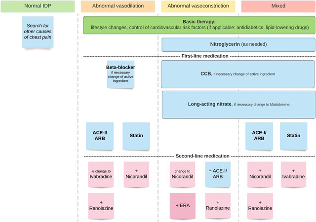
Figure 2. Endotypes to therapy based on the results of the interventional diagnostic procedure. IDP: Interventional diagnostic procedure; CCB: calcium channel blocker; ACE-I: angiotensin-converting enzyme inhibitor; ARB: angiotensin II receptor blocker; ERA: endothelin-receptor antagonist.
DECLARATIONS
Authors’ contributionsPerformed the literature search, data analysis and she produced the first draft of the manuscript: McChord J
Had the idea for the article and the topic and made critical revisions of the manuscript: Ong P
Performed literature search and made critical revisions: Hubert A
Made suggestions for the scope of the article and made critical revisions: Bekeredjian R
Availability of data and materialsNot applicable.
Financial support and sponsorshipThis work was supported by funds from the Berthold-Leibinger-Foundation, Ditzingen, Germany.
Conflicts of interestAll authors declared that there are no conflicts of interest.
Ethical approval and consent to participateNot applicable.
Consent for publicationNot applicable.
Copyright© The Author(s) 2021.
REFERENCES
2. Crea F, Camici PG, Bairey Merz CN. Coronary microvascular dysfunction: an update. Eur Heart J 2014;35:1101-11.
3. Murthy VL, Naya M, Taqueti VR, et al. Effects of sex on coronary microvascular dysfunction and cardiac outcomes. Circulation 2014;129:2518-27.
4. Gulati M, Cooper-DeHoff RM, McClure C, et al. Adverse cardiovascular outcomes in women with nonobstructive coronary artery disease: a report from the Women's Ischemia Syndrome Evaluation Study and the St James Women Take Heart Project. Arch Intern Med 2009;169:843-50.
5. Huang FY, Huang BT, Lv WY, et al. The prognosis of patients with nonobstructive coronary artery disease versus normal arteries determined by invasive coronary angiography or computed tomography coronary angiography: a systematic review. Medicine (Baltimore) 2016;95:e3117.
6. Johnson BD, Shaw LJ, Buchthal SD, et al. National Institutes of Health-National Heart, Lung, and Blood Institute. Prognosis in women with myocardial ischemia in the absence of obstructive coronary disease: results from the National Institutes of Health-National Heart, Lung, and Blood Institute-Sponsored Women's Ischemia Syndrome Evaluation (WISE). Circulation 2004;109:2993-9.
7. Lin FY, Shaw LJ, Dunning AM, et al. Mortality risk in symptomatic patients with nonobstructive coronary artery disease: a prospective 2-center study of 2,583 patients undergoing 64-detector row coronary computed tomographic angiography. J Am Coll Cardiol 2011;58:510-9.
8. Pepine CJ, Anderson RD, Sharaf BL, et al. Coronary microvascular reactivity to adenosine predicts adverse outcome in women evaluated for suspected ischemia results from the National Heart, Lung and Blood Institute WISE (Women's Ischemia Syndrome Evaluation) study. J Am Coll Cardiol 2010;55:2825-32.
9. Mohandas R, Segal MS, Huo T, et al. Renal function and coronary microvascular dysfunction in women with symptoms/signs of ischemia. PLoS One 2015;10:e0125374.
10. Kunadian V, Chieffo A, Camici PG, et al. An EAPCI expert consensus document on ischaemia with non-obstructive coronary arteries in Collaboration with European Society of Cardiology Working Group on Coronary Pathophysiology & Microcirculation Endorsed by Coronary Vasomotor Disorders International Study Group. Eur Heart J 2020;41:3504-20.
11. Camici PG, Olivotto I, Rimoldi OE. The coronary circulation and blood flow in left ventricular hypertrophy. J Mol Cell Cardiol 2012;52:857-64.
12. Ong P, Athanasiadis A, Borgulya G, Mahrholdt H, Kaski JC, Sechtem U. High prevalence of a pathological response to acetylcholine testing in patients with stable angina pectoris and unobstructed coronary arteries. The ACOVA Study (Abnormal COronary VAsomotion in patients with stable angina and unobstructed coronary arteries). J Am Coll Cardiol 2012;59:655-62.
13. Reis SE, Holubkov R, Conrad Smith AJ, et al. WISE Investigators. Coronary microvascular dysfunction is highly prevalent in women with chest pain in the absence of coronary artery disease: results from the NHLBI WISE study. Am Heart J 2001;141:735-41.
14. Wessel TR, Arant CB, McGorray SP, et al. NHLBI Women's Ischemia Syndrome Evaluation (WISE). Coronary microvascular reactivity is only partially predicted by atherosclerosis risk factors or coronary artery disease in women evaluated for suspected ischemia: results from the NHLBI Women's Ischemia Syndrome Evaluation (WISE). Clin Cardiol 2007;30:69-74.
15. Patel MR, Peterson ED, Dai D, et al. Low diagnostic yield of elective coronary angiography. N Engl J Med 2010;362:886-95.
16. Sara JD, Widmer RJ, Matsuzawa Y, Lennon RJ, Lerman LO, Lerman A. Prevalence of coronary microvascular dysfunction among patients with chest pain and nonobstructive coronary artery disease. JACC Cardiovasc Interv 2015;8:1445-53.
17. Lee BK, Lim HS, Fearon WF, et al. Invasive evaluation of patients with angina in the absence of obstructive coronary artery disease. Circulation 2015;131:1054-60.
18. Ford TJ, Stanley B, Good R, et al. Stratified medical therapy using invasive coronary function testing in angina: The CorMicA Trial. J Am Coll Cardiol 2018;72:2841-55.
19. Ong P, Safdar B, Seitz A, Hubert A, Beltrame JF, Prescott E. Diagnosis of coronary microvascular dysfunction in the clinic. Cardiovasc Res 2020;116:841-55.
20. Ong P, Camici PG, Beltrame JF, et al. Coronary Vasomotion Disorders International Study Group (COVADIS). International standardization of diagnostic criteria for microvascular angina. Int J Cardiol 2018;250:16-20.
21. Hoffman JI. Maximal coronary flow and the concept of coronary vascular reserve. Circulation 1984;70:153-9.
22. Gould K, Lipscomb K, Hamilton GW. Physiologic basis for assessing critical coronary stenosis. Am J Cardiol 1974;33:87-94.
23. Goodwill AG, Dick GM, Kiel AM, Tune JD. . Regulation of coronary blood flow. In: Terjung R, editor. Comprehensive physiology. Wiley; 2011. p. 321-82.
24. Bruyne B, Bartunek J, Sys SU, Pijls NH, Heyndrickx GR, Wijns W. Simultaneous coronary pressure and flow velocity measurements in humans. Feasibility, reproducibility, and hemodynamic dependence of coronary flow velocity reserve, hyperemic flow versus pressure slope index, and fractional flow reserve. Circulation 1996;94:1842-9.
25. Adjedj J, Toth GG, Johnson NP, et al. Intracoronary adenosine: dose-response relationship with hyperemia. JACC Cardiovasc Interv 2015;8:1422-30.
26. Granger DN, Rodrigues SF, Yildirim A, Senchenkova EY. Microvascular responses to cardiovascular risk factors. Microcirculation 2010;17:192-205.
27. Chahine RA, Feldman RL, Giles TD, et al. Randomized placebo-controlled trial of amlodipine in vasospastic angina. J Am Coll Cardiol 1993;21:1365-70.
28. Rosenthal SJ, Ginsburg R, Lamb IH, Baim DS, Schroeder JS. Efficacy of diltiazem for control of symptoms of coronary arterial spasm. Am J Cardiol 1980;46:1027-32.
29. Lanza GA, Colonna G, Pasceri V, Maseri A. Atenolol versus amlodipine versus isosorbide-5-mononitrate on anginal symptoms in syndrome X. Am J Cardiol 1999;84:854-6.
30. Masumoto A, Mohri M, Takeshita A. Three-year follow-up of the Japanese patients with microvascular angina attributable to coronary microvascular spasm. Int J Cardiol 2001;81:151-6.
31. Sorop O, Bakker EN, Pistea A, Spaan JA, VanBavel E. Calcium channel blockade prevents pressure-dependent inward remodeling in isolated subendocardial resistance vessels. Am J Physiol Heart Circ Physiol 2006;291:H1236-45.
32. Yao K, Nagashima K, Miki H. Pharmacological, pharmacokinetic, and clinical properties of benidipine hydrochloride, a novel, long-acting calcium channel blocker. J Pharmacol Sci 2006;100:243-61.
33. Abe M, Okada K, Maruyama N, et al. Benidipine reduces albuminuria and plasma aldosterone in mild-to-moderate stage chronic kidney disease with albuminuria. Hypertens Res 2011;34:268-73.
34. Tani S, Takahashi A, Nagao K, Hirayama A. Effects of the T/L-type calcium channel blocker benidipine on albuminuria and plasma aldosterone concentration. A pilot study involving switching from L-type calcium channel blockers to benidipine. Int Heart J 2014;55:519-25.
35. Unger T, Paulis L, Sica DA. Therapeutic perspectives in hypertension: novel means for renin-angiotensin-aldosterone system modulation and emerging device-based approaches. Eur Heart J 2011;32:2739-47.
36. Kosaka H, Hirayama K, Yoda N, et al. The L-, N-, and T-type triple calcium channel blocker benidipine acts as an antagonist of mineralocorticoid receptor, a member of nuclear receptor family. Eur J Pharmacol 2010;635:49-55.
37. Fuji Y, Suzuki H, Katsumata H, Nakajima S, Saruta T. Hormonal and renal responses to oral once-daily calcium entry blocker in normotensive and hypertensive persons. J Cardiovasc Pharmacol 1988;11:438-43.
38. Nishigaki K, Inoue Y, Yamanouchi Y, et al. Prognostic effects of calcium channel blockers in patients with vasospastic angina--a meta-analysis. Circ J 2010;74:1943-50.
39. Kim SE, Jo SH, Han SH, et al. Comparison of calcium-channel blockers for long-term clinical outcomes in patients with vasospastic angina. Korean J Intern Med 2021;36:124-34.
40. Miwa Y, Masai H, Shimizu M. Differential effects of calcium-channel blockers on vascular endothelial function in patients with coronary spastic angina. Circ J 2009;73:713-7.
41. Day L, Sowton E. Clinical features and follow-up of patients with angina and normal coronary arteries. Lancet 1976;308:334-7.
42. Isner JM, Fisher GP, Del Negro AA, Borer JS. Right ventricular infarction with hemodynamic decompensation due to transient loss of active atrial augmentation: successful treatment with atrial pacing. Am Heart J 1981;102:792-4.
43. Matsumoto T, Takahashi M, Omura T, et al. Heterogeneity in the vasorelaxing effect of nicorandil on dog epicardial coronary arteries: comparison with other NO donors. J Cardiovasc Pharmacol 1997;29:772-9.
44. Kim CH, Park TK, Cho SW, et al. Impact of different nitrate therapies on long-term clinical outcomes of patients with vasospastic angina: a propensity score-matched analysis. Int J Cardiol 2018;252:1-5.
45. Kukovetz WR, Holzmann S, Braida C, Pöch G. Dual mechanism of the relaxing effect of nicorandil by stimulation of cyclic GMP formation and by hyperpolarization. J Cardiovasc Pharmacol 1991;17:627-33.
46. Kukovetz WR, Holzmann S, Pöch G. Molecular mechanism of action of nicorandil. J Cardiovasc Pharmacol 1992;20:S1-7.
47. Tarkin JM, Kaski JC. Vasodilator therapy: nitrates and nicorandil. Cardiovasc Drugs Ther 2016;30:367-78.
48. Brodmann M, Lischnig U, Lueger A, Stark G, Pilger E. The effect of the K+ agonist nicorandil on peripheral vascular resistance. Int J Cardiol 2006;111:49-52.
49. Kool MJ, Spek JJ, Struyker Boudier HA, et al. Acute and subacute effects of nicorandil and isosorbide dinitrate on vessel wall properties of large arteries and hemodynamics in healthy volunteers. Cardiovasc Drugs Ther 1995;9:331-7.
50. Ishibashi Y, Takahashi N, Tokumaru A, et al. Effects of long-term nicorandil administration on endothelial function, inflammation, and oxidative stress in patients without coronary artery disease. J Cardiovasc Pharmacol 2008;51:311-6.
51. Lindemann H, Petrovic I, Hill S, et al. Biopsy-confirmed endothelial cell activation in patients with coronary microvascular dysfunction. Coron Artery Dis 2018;29:216-22.
52. Frishman WH. β-Adrenergic blockade in cardiovascular disease. J Cardiovasc Pharmacol Ther 2013;18:310-9.
53. Duncker DJ, Koller A, Merkus D, Canty JM Jr. Regulation of coronary blood flow in health and ischemic heart disease. Prog Cardiovasc Dis 2015;57:409-22.
54. Kalinowski L, Dobrucki LW, Szczepanska-Konkel M, et al. Third-generation beta-blockers stimulate nitric oxide release from endothelial cells through ATP efflux: a novel mechanism for antihypertensive action. Circulation 2003;107:2747-52.
55. Mason RP, Jacob RF, Corbalan JJ, Szczesny D, Matysiak K, Malinski T. The favorable kinetics and balance of nebivolol-stimulated nitric oxide and peroxynitrite release in human endothelial cells. BMC Pharmacol Toxicol 2013;14:48.
56. Shahin Y, Khan JA, Samuel N, Chetter I. Angiotensin converting enzyme inhibitors effect on endothelial dysfunction: a meta-analysis of randomised controlled trials. Atherosclerosis 2011;216:7-16.
57. Büchner N, Banas B, Krämer BK. Telmisartan, ramipril, or both in patients at high risk of vascular events. N Engl J Med 2008;359:426.
58. Dagenais GR, Yusuf S, Bourassa MG, et al. HOPE Investigators. Effects of ramipril on coronary events in high-risk persons: results of the Heart Outcomes Prevention Evaluation Study. Circulation 2001;104:522-6.
59. Pauly DF, Johnson BD, Anderson RD, et al. In women with symptoms of cardiac ischemia, nonobstructive coronary arteries, and microvascular dysfunction, angiotensin-converting enzyme inhibition is associated with improved microvascular function: A double-blind randomized study from the National Heart, Lung and Blood Institute Women's Ischemia Syndrome Evaluation (WISE). Am Heart J 2011;162:678-84.
60. Suhrs HE, Michelsen MM, Prescott E. Treatment strategies in coronary microvascular dysfunction: a systematic review of interventional studies. Microcirculation 2019;26:e12430.
61. Lefer A. Vascular effects of HMG CoA-reductase inhibitors (statins) unrelated to cholesterol lowering: new concepts for cardiovascular disease. Cardiovasc Res 2001;49:281-7.
62. Rosenson RS. Statin therapy: new therapy for cardiac microvascular dysfunction. Eur Heart J 2003;24:1993-4.
63. Zhang X, Li Q, Zhao J, et al. Effects of combination of statin and calcium channel blocker in patients with cardiac syndrome X. Coron Artery Dis 2014;25:40-4.
64. Yokoyama I, Inoue Y, Moritan T, Ohtomo K, Nagai R. Impaired myocardial vasodilatation during hyperaemic stress is improved by simvastatin but not by pravastatin in patients with hypercholesterolaemia. Eur Heart J 2004;25:671-9.
65. Jia Q, Shi S, Yuan G, et al. The effect of nicorandil in patients with cardiac syndrome X: a meta-analysis of randomized controlled trials. Medicine (Baltimore) 2020;99:e22167.
66. Bassenge E, Pohl U. Effect of molsidomine on cardiac preload, coronary artery diameter, and coronary resistance. Am Heart J 1985;109:627-30.
67. Tagliamonte E, Rigo F, Cirillo T, et al. Effects of ranolazine on noninvasive coronary flow reserve in patients with myocardial ischemia but without obstructive coronary artery disease. Echocardiography 2015;32:516-21.
68. D'Elia E, Fiocca L, Ferrero P, et al. Ranolazine in heart failure with preserved left ventricular ejection fraction and microvascular dysfunction: case report and literature review. J Clin Pharmacol 2013;53:665-9.
69. Cattaneo M, Porretta AP, Gallino A. Ranolazine: Drug overview and possible role in primary microvascular angina management. Int J Cardiol 2015;181:376-81.
70. Sharp RP, Patatanian E, Sirajuddin R. Use of ranolazine for the treatment of coronary microvascular dysfunction. Am J Cardiovasc Drugs 2021; doi: 10.1007/s40256-020-00462-6.
71. DiFrancesco D. Characterization of single pacemaker channels in cardiac sino-atrial node cells. Nature 1986;324:470-3.
72. DiFrancesco D. The contribution of the 'pacemaker' current (if) to generation of spontaneous activity in rabbit sino-atrial node myocytes. J Physiol 1991;434:23-40.
73. Borer JS, Fox K, Jaillon P, Lerebours G. Ivabradine Investigators Group. Antianginal and antiischemic effects of ivabradine, an I(f) inhibitor, in stable angina: a randomized, double-blind, multicentered, placebo-controlled trial. Circulation 2003;107:817-23.
74. Villano A, Di Franco A, Nerla R, et al. Effects of ivabradine and ranolazine in patients with microvascular angina pectoris. Am J Cardiol 2013;112:8-13.
75. Skalidis EI, Hamilos MI, Chlouverakis G, Zacharis EA, Vardas PE. Ivabradine improves coronary flow reserve in patients with stable coronary artery disease. Atherosclerosis 2011;215:160-5.
76. Camici PG, Gloekler S, Levy BI, et al. Ivabradine in chronic stable angina: effects by and beyond heart rate reduction. Int J Cardiol 2016;215:1-6.
77. Maseri A, Crea F, Kaski JC, Crake T. Mechanisms of angina pectoris in syndrome X. J Am Coll Cardiol 1991;17:499-506.
78. Lanza GA, Crea F. Primary coronary microvascular dysfunction: clinical presentation, pathophysiology, and management. Circulation 2010;121:2317-25.
79. Emdin M, Picano E, Lattanzi F, l'Abbate A. Improved exercise capacity with acute aminophylline administration in patients with syndrome X. J Am Coll Cardiol 1989;14:1450-3.
80. Ferrari R, Camici PG, Crea F, et al. Expert consensus document: a 'diamond' approach to personalized treatment of angina. Nat Rev Cardiol 2018;15:120-32.
81. Yasaei R, Peterson E, Saadabadi A. . StatPearls: Chronic Pain Syndrome. In: StatPearls [Internet]. Treasure Island (FL): StatPearls Publishing; 2021.
82. Sanderson JE, Brooksby P, Waterhouse D, Palmer RB, Neubauer K. Epidural spinal electrical stimulation for severe angina: a study of its effects on symptoms, exercise tolerance and degree of ischaemia. Eur Heart J 1992;13:628-33.
83. Lanza GA, Sestito A, Sgueglia GA, et al. Effect of spinal cord stimulation on spontaneous and stress-induced angina and 'ischemia-like' ST-segment depression in patients with cardiac syndrome X. Eur Heart J 2005;26:983-9.
84. Mannheimer C, Eliasson T, Augustinsson LE, et al. Electrical stimulation versus coronary artery bypass surgery in severe angina pectoris: the ESBY study. Circulation 1998;97:1157-63.
85. Sgueglia GA, Sestito A, Spinelli A, et al. Long-term follow-up of patients with cardiac syndrome X treated by spinal cord stimulation. Heart 2007;93:591-7.
86. Arora RR, Chou TM, Jain D, et al. Effects of enhanced external counterpulsation on Health-Related Quality of Life continue 12 months after treatment: a substudy of the Multicenter Study of Enhanced External Counterpulsation. J Investig Med 2002;50:25-32.
87. Arora RR, Chou TM, Jain D, et al. The multicenter study of enhanced external counterpulsation (MUST-EECP): effect of EECP on exercise-induced myocardial ischemia and anginal episodes. J Am Coll Cardiol 1999;33:1833-40.
88. Maccarthy PA, Pegge NC, Prendergast BD, Shah AM, Groves PH. The physiological role of endogenous endothelin in the regulation of human coronary vasomotor tone. J Am Coll Cardiol 2001;37:137-43.
89. Mather KJ, Lteif AA, Veeneman E, et al. Role of endogenous ET-1 in the regulation of myocardial blood flow in lean and obese humans. Obesity (Silver Spring) 2010;18:63-70.
90. Kaski JC, Elliott PM, Salomone O, et al. Concentration of circulating plasma endothelin in patients with angina and normal coronary angiograms. Br Heart J 1995;74:620-4.
91. Cox ID, Bøtker HE, Bagger JP, Sonne HS, Kristensen BØ, Kaski JC. Elevated endothelin concentrations are associated with reduced coronary vasomotor responses in patients with chest pain and normal coronary arteriograms. J Am Coll Cardiol 1999;34:455-60.
92. Johnson NP, Gould KL. Physiology of endothelin in producing myocardial perfusion heterogeneity: a mechanistic study using darusentan and positron emission tomography. J Nucl Cardiol 2013;20:835-44.
93. Reriani M, Raichlin E, Prasad A, et al. Long-term administration of endothelin receptor antagonist improves coronary endothelial function in patients with early atherosclerosis. Circulation 2010;122:958-66.
94. Ford TJ, Rocchiccioli P, Good R, et al. Systemic microvascular dysfunction in microvascular and vasospastic angina. Eur Heart J 2018;39:4086-97.
95. Greene SJ, Gheorghiade M, Borlaug BA, et al. The cGMP signaling pathway as a therapeutic target in heart failure with preserved ejection fraction. J Am Heart Assoc 2013;2:e000536.
96. Follmann M, Ackerstaff J, Redlich G, et al. Discovery of the soluble guanylate cyclase stimulator vericiguat (BAY 1021189) for the treatment of chronic heart failure. J Med Chem 2017;60:5146-61.
97. Martínez Pereyra V, Seitz A, Hubert A, et al. Repurposing riociguat for treatment of refractory angina resulting from coronary spasm. JACC: Case Reports 2021;3:392-6.
Cite This Article
Export citation file: BibTeX | RIS
OAE Style
McChord J, Hubert A, Bekeredjian R, Ong P. Contemporary pharmacological treatment strategies for patients with angina and unobstructed coronary arteries (ANOCA) due to coronary microvascular dysfunction. Vessel Plus 2021;5:49. http://dx.doi.org/10.20517/2574-1209.2021.63
AMA Style
McChord J, Hubert A, Bekeredjian R, Ong P. Contemporary pharmacological treatment strategies for patients with angina and unobstructed coronary arteries (ANOCA) due to coronary microvascular dysfunction. Vessel Plus. 2021; 5: 49. http://dx.doi.org/10.20517/2574-1209.2021.63
Chicago/Turabian Style
McChord, Johanna, Astrid Hubert, Raffi Bekeredjian, Peter Ong. 2021. "Contemporary pharmacological treatment strategies for patients with angina and unobstructed coronary arteries (ANOCA) due to coronary microvascular dysfunction" Vessel Plus. 5: 49. http://dx.doi.org/10.20517/2574-1209.2021.63
ACS Style
McChord, J.; Hubert A.; Bekeredjian R.; Ong P. Contemporary pharmacological treatment strategies for patients with angina and unobstructed coronary arteries (ANOCA) due to coronary microvascular dysfunction. Vessel Plus. 2021, 5, 49. http://dx.doi.org/10.20517/2574-1209.2021.63
About This Article
Copyright

Data & Comments
Data


 Cite This Article 8 clicks

 Like This Article 0
likes
Like This Article 0
likes


Comments
Comments must be written in English. Spam, offensive content, impersonation, and private information will not be permitted. If any comment is reported and identified as inappropriate content by OAE staff, the comment will be removed without notice. If you have any queries or need any help, please contact us at support@oaepublish.com.